
Posterior Collection (#6)

"Exploring the Fascinating Posterior

Background imagePosterior Collection: Calf muscles, artwork

Calf muscles, artwork
Calf muscles. Computer artwork of a posterior view of some of the muscles and tendons of the calves and their attachment to the bones of the leg

Background imagePosterior Collection: Back muscles, anatomical artwork

Back muscles, anatomical artwork
Back muscles. Anatomical computer artwork showing the muscles of the human back, and shoulders

Background imagePosterior Collection: Knee ligaments, artwork

Knee ligaments, artwork
Knee ligaments. Computer artwork of a human knee joint, showing the bones (white) and ligaments (purple). The knee joint is formed by the articulation of the femur (thigh bone)

Background imagePosterior Collection: Upper back anatomy, artwork

Upper back anatomy, artwork
Upper back anatomy, computer artwork. The muscles either side of the neck that attach to the shoulder blades (scapulae) are the levator scapulae muscles

Background imagePosterior Collection: Nuchal ligament, artwork

Nuchal ligament, artwork
Nuchal ligament. Computer artwork showing the bones of the neck and head, and the nuchal ligament (white, down centre of spine). Ligaments are bands of connective tissue that connect bones

Background imagePosterior Collection: Suboccipital muscles and nerve, artwork C014 / 5099

Suboccipital muscles and nerve, artwork C014 / 5099
Suboccipital muscles. Computer artwork of the back of the base of the skull showing nerves (yellow) and the suboccipital muscles (pink)

Background imagePosterior Collection: Suboccipital muscles and nerve, artwork C014 / 5098

Suboccipital muscles and nerve, artwork C014 / 5098
Suboccipital muscles. Computer artwork of the back of the base of the skull showing nerves (yellow) and the suboccipital muscles (pink)

Background imagePosterior Collection: Human muscle anatomy, 19th century C015 / 6103

Human muscle anatomy, 19th century C015 / 6103
Human muscle anatomy. 19th-century artwork of a posterior view of the first and second layers of human musculature. Artwork from Encyclopaedia Metropolitana

Background imagePosterior Collection: Calf muscles, artwork C014 / 5063

Calf muscles, artwork C014 / 5063
Calf muscles. Computer artwork of a posterior view of some of the muscles and tendons of the calves and their attachment to the bones of the leg

Background imagePosterior Collection: Abdominal external oblique muscle C014 / 5062

Abdominal external oblique muscle C014 / 5062
Abdominal external oblique muscle, computer artwork. This thin muscle acts to pull the chest down and compress the abdominal cavity

Background imagePosterior Collection: Heart and lung anatomy, artwork C016 / 2904

Heart and lung anatomy, artwork C016 / 2904
Heart and lung anatomy. Artwork of a posterior (from behind) view of the heart and lungs. The heart (mostly obscured) is a hollow muscle that pumps blood around the body and to the lungs

Background imagePosterior Collection: Single-kidney anatomy, artwork C016 / 2903

Single-kidney anatomy, artwork C016 / 2903
Single-kidney anatomy. Artwork showing a posterior (from behind) view of human kidneys in relation to the main torso blood vessels (vena cava, blue; aorta, red) and the male abdomen

Background imagePosterior Collection: Single-kidney anatomy, artwork C016 / 2897

Single-kidney anatomy, artwork C016 / 2897
Single-kidney anatomy. Artwork showing a posterior (from behind) view of human kidneys in relation to the bones of the skeleton. One kidney (brown, upper right) has a normal appearance

Background imagePosterior Collection: Knee ligaments, artwork C013 / 4662

Knee ligaments, artwork C013 / 4662
Knee ligaments. Computer artwork of a posterior view of the knee (left), showing some of the ligaments (white), membranes and other connective tissues forming this joint

Background imagePosterior Collection: Collateral knee ligaments, artwork

Collateral knee ligaments, artwork
Knee ligaments. Computer artwork of a posterior view of the knee, showing some of the ligaments (white), membranes and other connective tissues forming this joint

Background imagePosterior Collection: Calf muscles, artwork C013 / 4576

Calf muscles, artwork C013 / 4576
Calf muscles. Computer artwork of a posterior view of a set of calf muscles (red) and their attachments to the bones of the leg and foot

Background imagePosterior Collection: Calf muscles, artwork C013 / 4572

Calf muscles, artwork C013 / 4572
Calf muscles. Computer artwork of an oblique posterior view of some of the muscles and tendons (red and white) of the calves and their attachment to the bones of the leg

Background imagePosterior Collection: Calf muscles, artwork C013 / 4571

Calf muscles, artwork C013 / 4571
Calf muscles. Computer artwork of an oblique posterior view of some of the muscles and tendons (red and white) of the calves and their attachment to the bones of the leg

Background imagePosterior Collection: Chest muscles, artwork C013 / 4566

Chest muscles, artwork C013 / 4566
Chest muscles. Computer artwork of an oblique posterior view from above of some of the muscles (red) of the chest, showing their attachment to the bones of the ribcage and back

Background imagePosterior Collection: Shoulder muscles, artwork C013 / 4515

Shoulder muscles, artwork C013 / 4515
Shoulder muscles. Computer artwork of a posterior view of two of the muscles (red) of the shoulders and their attachment to the bones of the back and upper arms

Background imagePosterior Collection: Shoulder muscle, artwork C013 / 4508

Shoulder muscle, artwork C013 / 4508
Shoulder muscle. Computer artwork of an oblique posterior view from below of one of the muscles (red) of the shoulders and its attachment to the bones of the back and upper arms

Background imagePosterior Collection: Shoulder muscle, artwork C013 / 4507

Shoulder muscle, artwork C013 / 4507
Shoulder muscle. Computer artwork of an oblique posterior view from above of one of the muscles (red) of the shoulders and its attachment to the bones of the back and upper arms

Background imagePosterior Collection: Leg anatomy, artwork C013 / 4497

Leg anatomy, artwork C013 / 4497
Leg anatomy. Computer artwork of an oblique posterior view looking up a human leg, showing muscles and tendons (brown and white), bones and ligaments (white), arteries and veins (red and blue)

Background imagePosterior Collection: Foot muscles, artwork C013 / 4483

Foot muscles, artwork C013 / 4483
Foot muscles. Computer artwork of an oblique view from below and behind of some of the muscles and tendons (red and white) of the sole of the foot

Background imagePosterior Collection: Foot muscles, artwork C013 / 4478

Foot muscles, artwork C013 / 4478
Foot muscles. Computer artwork of a view from below of some of the muscles and tendons (red and white) of the sole of the foot

Background imagePosterior Collection: Foot muscles, artwork C013 / 4477

Foot muscles, artwork C013 / 4477
Foot muscles. Computer artwork of a view from below of some of the muscles and tendons (red and white) of the sole of the foot

Background imagePosterior Collection: Hip ligaments, artwork C013 / 4434

Hip ligaments, artwork C013 / 4434
Hip ligaments. Computer artwork of a posterior oblique view of the hip bones and ligaments (white). At upper right the lumbar spine ends in the sacrum and coccyx (tailbone)

Background imagePosterior Collection: Hip ligaments, artwork C013 / 4433

Hip ligaments, artwork C013 / 4433
Hip ligaments. Computer artwork of a posterior view of the hip bones and ligaments (white). At centre the lumbar spine ends in the sacrum and coccyx (tailbone)

Background imagePosterior Collection: Leg muscles, artwork C013 / 4429

Leg muscles, artwork C013 / 4429
Leg muscles. Computer artwork of the bones and outer muscles of the left leg, seen from the front (anterior view) at an oblique angle looking downwards

Background imagePosterior Collection: Thigh hamstring muscles, artwork C013 / 4428

Thigh hamstring muscles, artwork C013 / 4428
Thigh hamstring muscles. Computer artwork of a posterior view of two of the three hamstring muscles (red) of the thighs. The muscles are the semimembranosus (inner) and semitendinosus (outer)

Background imagePosterior Collection: Buttock muscles, artwork C013 / 4423

Buttock muscles, artwork C013 / 4423
Buttock muscles. Computer artwork of a posterior oblique view of the hips, showing some of the muscles (red) of the buttocks

Background imagePosterior Collection: Buttock muscles, artwork C013 / 4424

Buttock muscles, artwork C013 / 4424
Buttock muscles. Computer artwork of a posterior view of the hips showing some of the muscles (red) of the buttocks. These are shown attached to the femurs (thigh bones) and the pelvis bones

Background imagePosterior Collection: Buttock muscles, artwork C013 / 4422

Buttock muscles, artwork C013 / 4422
Buttock muscles. Computer artwork of a posterior oblique view of the hips showing some of the muscles (red) of the buttocks. These are shown attached to the femurs (thigh bones) and the pelvis bones

Background imagePosterior Collection: Buttock muscles, artwork C013 / 4420

Buttock muscles, artwork C013 / 4420
Buttock muscles. Computer artwork of a posterior view of the hips showing some of the muscles (red) of the buttocks. These are shown attached to the femurs (thigh bones) and the pelvis bones

Background imagePosterior Collection: Buttock muscles, artwork C013 / 4419

Buttock muscles, artwork C013 / 4419
Buttock muscles. Computer artwork of a posterior oblique view of the hips showing some of the muscles (red) of the buttocks. These are shown attached to the femurs (thigh bones) and the pelvis bones

Background imagePosterior Collection: Buttock muscles, artwork C013 / 4415

Buttock muscles, artwork C013 / 4415
Buttock muscles. Computer artwork of a posterior oblique view of the hips showing some of the muscles (red) of the buttocks. These are shown attached to the femurs (thigh bones) and the pelvis bones

Background imagePosterior Collection: Buttock muscles, artwork C013 / 4416

Buttock muscles, artwork C013 / 4416
Buttock muscles. Computer artwork of a posterior oblique view of the hips showing some of the muscles (red) of the buttocks. These are shown attached to the femurs (thigh bones) and the pelvis bones

Background imagePosterior Collection: An Agouti (Dasyprocta punctata) eats a seed pod on a remote beach with in Corcovado National Park

An Agouti (Dasyprocta punctata) eats a seed pod on a remote beach with in Corcovado National Park, Costa Rica

Background imagePosterior Collection: Eurasian Linnet (Carduelis cannabina) adult pair, mating, female showing cloaca

Eurasian Linnet (Carduelis cannabina) adult pair, mating, female showing cloaca, perched on flowering gorse, Norfolk, England, april

Background imagePosterior Collection: Indian rhinoceros

Indian rhinoceros (Rhinoceros unicornis) grazing. The Indian rhinoceros is found in Nepal and Assam, India. It can run up to speeds of 34 miles per hour (mph)

Background imagePosterior Collection: Larger grain borer beetle, SEM

Larger grain borer beetle, SEM
Larger grain borer. Coloured scanning electron micrograph (SEM) of a larger grain borer beetle (Prostephanus truncatus) boring into a kernel of maize

Background imagePosterior Collection: Neck and skull, X-ray

Neck and skull, X-ray
Neck and skull. Coloured X-ray of a normal human neck (lower centre) and part of the skull (upper centre). The neck joins the head to the body

Background imagePosterior Collection: Fractured atlas vertebra, 3D CT scan

Fractured atlas vertebra, 3D CT scan
Fractured atlas vertebra, coloured 3D CT (computed tomography) scan. Posterior view of a fractured atlas vertebra (break at upper centre) of a 34 year old man. Part of the skull can be seen at top

Background imagePosterior Collection: Heart and lungs

Heart and lungs. Historical artwork of the heart (pink) and lungs (red, left and right) seen from behind. Dissection hooks have been used to draw back the lung tissue

Background imagePosterior Collection: Leg nerves

Leg nerves. Historical anatomical artwork of the nerves (white) of the rear of a human lower leg. At left, the superficial (surface) nerves are seen and at right the deep nerves and muscles (red)

Background imagePosterior Collection: Knee bones and ligaments

Knee bones and ligaments. Historical anatomical artwork of knee bones (yellow) and ligaments (pale blue). Ligaments are bands of fibrous tissue that hold bones together at joints

Background imagePosterior Collection: Hand bones and ligaments

Hand bones and ligaments. Historical anatomical artwork of the bones (yellow) & ligaments (white) of the hand. Ligaments are bands of fibrous tissue that hold bones together at their joints

Background imagePosterior Collection: Ankle bones and ligaments

Ankle bones and ligaments. Historical anatomical artwork of ankle bones (yellow) and ligaments (pale blue). Ligaments are bands of fibrous tissue that hold bones together at their joints


For sale as Licensed Images

Choose your image, Select your licence and Download the media

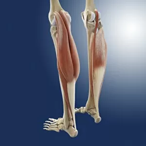
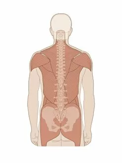
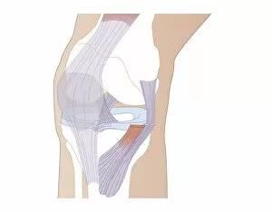
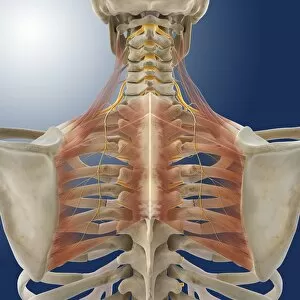
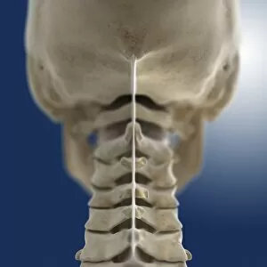
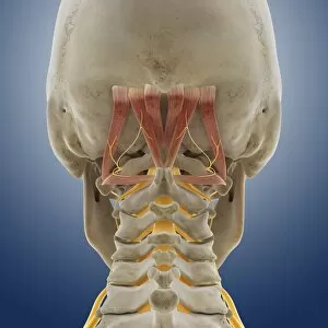
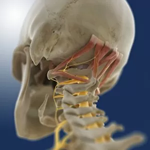
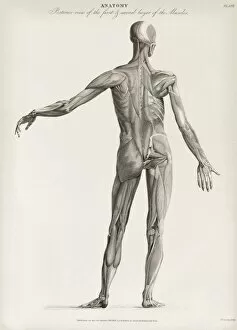
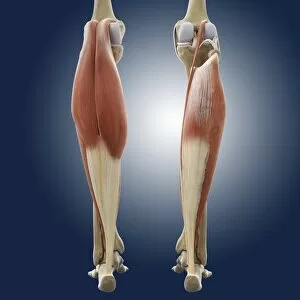
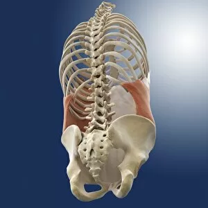
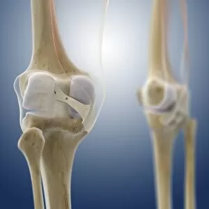
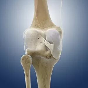
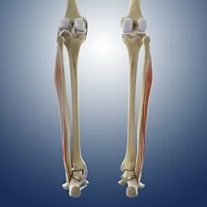
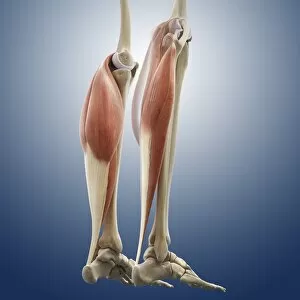
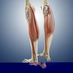
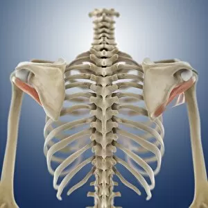
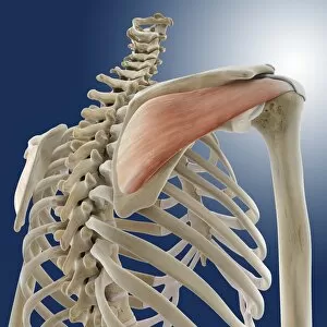
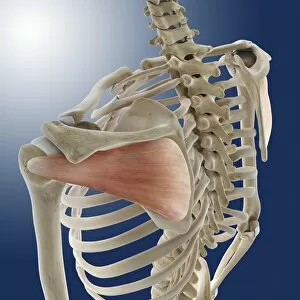
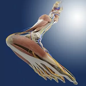
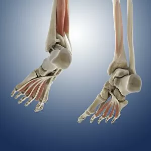
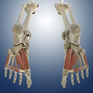
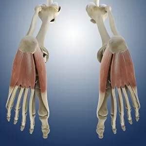
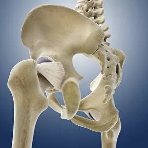
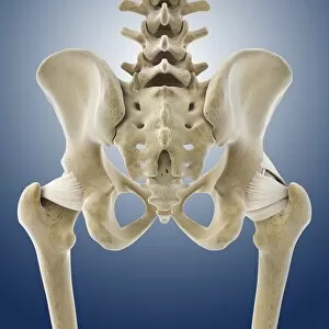
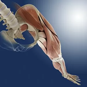
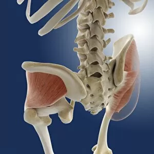
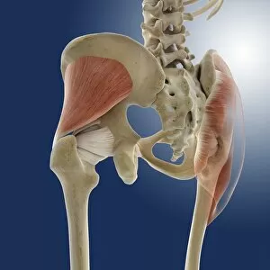
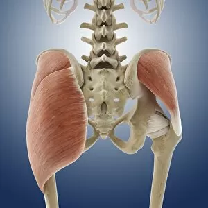
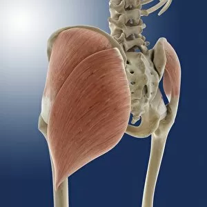
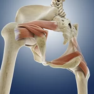
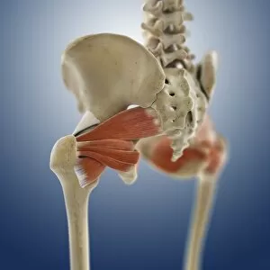
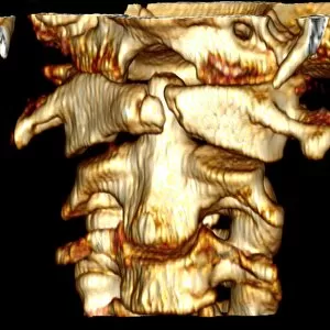
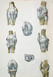
"Exploring the Fascinating Posterior: A Journey through Human Anatomy and Art" Step into a world where science meets art as we delve into the intricate details of the posterior. From Julien Bougle's captivating artwork to detailed anatomical studies, prepare to be amazed by the wonders that lie behind us. Julien Bougle's masterpiece showcases the human body with superimposed colored plates, highlighting its complexity and beauty. As we move closer, our focus shifts towards skull anatomy, where every bone tells a story of our past. The four views of the heart remind us of its vital role in sustaining life, while also showcasing its elegance from different angles. The seated male model immortalized on millboard captures both strength and vulnerability, allowing us to appreciate the intricacies of muscle structure. But let's not forget about one area that often goes unnoticed – the gluteal muscles in our buttocks play an essential role in our daily movements. As history unfolds before our eyes with "The Ides of March, " we witness how even ancient civilizations recognized the significance of this region. Moving deeper within ourselves, we explore engravings depicting roots of lungs and posterior pulmonary plexus seen from behind – a reminder that there is always more than meets the eye. Intriguingly detailed pen & ink studies reveal male nudes after 1501-2, capturing both vulnerability and strength simultaneously. And just when you think you've seen it all, a pair of broad bottoms depicted in vibrant colors reminds us that art has no boundaries when it comes to exploring every aspect of humanity. So join us on this captivating journey through time and space as we unravel secrets hidden within our own bodies' posterior regions. Let curiosity guide you as you discover new perspectives on what lies beneath - for there is much more than meets the eye when it comes to understanding ourselves fully.