
Rectum Collection (#5)
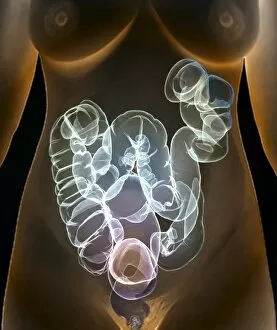
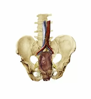
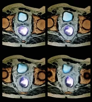
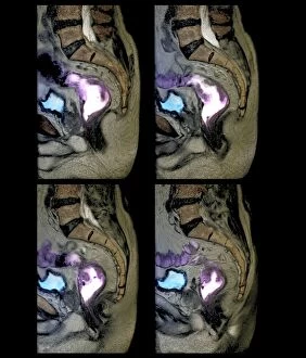
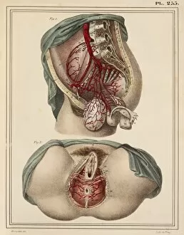
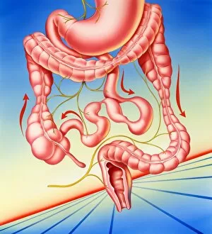
The intricate network of male groin arteries and the delicate balance within the human digestive system are depicted in this captivating 1825 artwork


For sale as Licensed Images
Choose your image, Select your licence and Download the media
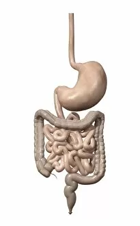
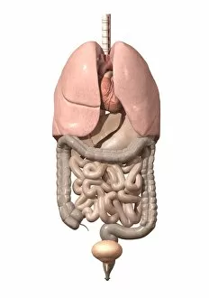
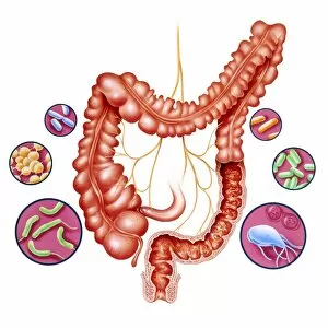
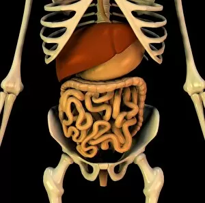
The intricate network of male groin arteries and the delicate balance within the human digestive system are depicted in this captivating 1825 artwork. From gastric bypass procedures to cross-sectional biomedical illustrations, every detail of the internal organs is meticulously portrayed. Explore the fascinating world of rectal exams, as seen in Curtis British Entomology Plate 538 and Ms 22532 Examination of a patient with hemorrhoids. Delve into ancient medical practices with Ms Arabe 3467 A Medical Examination from The Book of Kalila and Dimna, showcasing vellum-bound knowledge passed down through generations. Witness the complexity of normal male cross-section anatomy through digital illustrations and ultrasound technology-assisted rectal exams. Discover how these examinations provide valuable insights into reproductive health while also shedding light on other interconnected systems like respiration. As we unravel the mysteries hidden beneath our skin, let us not forget that even creatures like frogs have their own unique anatomical structures worth exploring. Join us on this journey through time and science as we delve deep into the wonders of the rectum.