Red Blood Cells Collection (#4)

"Exploring the Intricate World of Red Blood Cells: From Menstruation to Malaria" Uterus lining during menstruation

Background imageRed Blood Cells Collection: Sickle cell anaemia, light micrograph C015 / 6395

Sickle cell anaemia, light micrograph C015 / 6395
Sickle cell anaemia. Light micrograph of a blood smear showing normally rounded red blood cells that have become curved, elongated sickle-shaped cells

Background imageRed Blood Cells Collection: Trypanosomes in blood smear, SEM C016 / 5783

Trypanosomes in blood smear, SEM C016 / 5783
Parasitic protozoan. Coloured scanning electron micrograph (SEM) of trypanosome protozoa (Trypanosoma sp.) in a blood smear

Background imageRed Blood Cells Collection: Erythropoietin hormone complex

Erythropoietin hormone complex. Computer model showing the secondary structure of a molecule of the human hormone Erythropoietin (EPO), complexed with an erythropoetin receptor molecule

Background imageRed Blood Cells Collection: Blood clot C013 / 7129

Blood clot C013 / 7129
Blood clot. Coloured scanning electron micrograph (SEM) of a blood clot (centre) forming in connective tissue. Red blood cells (erythrocytes)

Background imageRed Blood Cells Collection: Blood clot C013 / 7128

Blood clot C013 / 7128
Blood clot. Coloured scanning electron micrograph (SEM) of a blood clot (centre) forming in connective tissue. Red blood cells (erythrocytes)

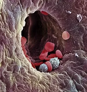
Background imageRed Blood Cells Collection: Blood-filled artery, SEM C013 / 7117

Blood-filled artery, SEM C013 / 7117
Blood-filled artery. Coloured scanning electron micrograph (SEM) of a section through an artery (dark brown) in muscle tissue (light brown), showing the red blood cells (erythrocytes, centre) inside

Background imageRed Blood Cells Collection: Blood-filled artery, SEM C013 / 7118

Blood-filled artery, SEM C013 / 7118
Blood-filled artery. Coloured scanning electron micrograph (SEM) of a section through an artery (centre) in muscle tissue, showing the red blood cells (erythrocytes, red) inside

Background imageRed Blood Cells Collection: Blood-filled artery, SEM C013 / 7116

Blood-filled artery, SEM C013 / 7116
Blood-filled artery. Coloured scanning electron micrograph (SEM) of a section through an artery (dark pink, centre) in muscle tissue (purple), showing the red blood cells (erythrocytes, red) inside

Background imageRed Blood Cells Collection: Blood cancer, conceptual image C013 / 4618

Blood cancer, conceptual image C013 / 4618
Blood cancer, conceptual image. Computer artwork of the interior of a blood vessel, showing cancer cells (dark) amongst normal red blood cells (red)

Background imageRed Blood Cells Collection: Blood cancer, conceptual image C013 / 4614

Blood cancer, conceptual image C013 / 4614
Blood cancer, conceptual image. Computer artwork of the interior of a blood vessel, showing cancer cells (white) amongst normal red blood cells (red)

Background imageRed Blood Cells Collection: Liver capillary, SEM

Liver capillary, SEM
Liver capillary. Coloured scanning electron micro- graph (SEM) of a freeze-fractured section through liver tissue showing a hepatic capillary, or sinusoid

Background imageRed Blood Cells Collection: Collapsed ovarian follicle, SEM

Collapsed ovarian follicle, SEM
Collapsed ovarian follicle. Coloured scanning electron micrograph (SEM) of an ovarian follicle just after ovulation (release of an egg)

Background imageRed Blood Cells Collection: Lung alveoli, SEM

Lung alveoli, SEM
Lung alveoli. Coloured scanning electron micrograph (SEM) of alveoli (air spaces) in the lungs. Blood vessels containing erythrocytes (red blood cells) are also seen

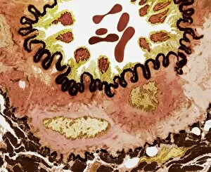
Background imageRed Blood Cells Collection: Contracted artery, TEM

Contracted artery, TEM
Contracted elastic artery. Coloured transmission electron micrograph (TEM) of a section through an artery. Red blood cells (erythrocytes) are seen in the lumen (top centre)

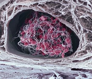
Background imageRed Blood Cells Collection: Blood clots, SEM

Blood clots, SEM
Blood clots. Coloured scanning electron micrograph (SEM) of small blood clots (red) in a blood vessel. The clots consist of red blood cells, white blood cells, and platelets

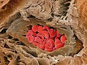
Background imageRed Blood Cells Collection: Coronary artery, SEM

Coronary artery, SEM
Coronary artery. Coloured scanning electron micrograph (SEM) of an artery in cardiac (heart) muscle. Red blood cells (erythrocytes) are seen within the vessel

Background imageRed Blood Cells Collection: Blood vessel types, artwork

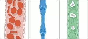
Blood vessel types, artwork
Blood vessel types. Artworks of the three types of blood vessels, from left: arteries, veins, and capillaries. Arteries are thick blood vessels carrying oxygenated blood around the body away from

Background imageRed Blood Cells Collection: Blood vessels, computer artwork

Blood vessels, computer artwork
Blood vessels. Computer artwork of two blood vessels. Red blood cells (erythrocytes) are seen within the blood vessels. The red blood cells distribute oxygen to body tissues

Background imageRed Blood Cells Collection: Ovarian blood vessel, SEM

Ovarian blood vessel, SEM
Ovarian blood vessel. Coloured scanning electron micrograph (SEM) of a section through a blood vessel in an ovary. Red blood cells are seen in the lumen, which is lined with endothelial cells (brown)

Background imageRed Blood Cells Collection: Nanorobots, computer artwork

Nanorobots, computer artwork. Nanotechnology is an area of science concerned with producing mechanical entities on the scale of nanometres (billionths of a metre)

Background imageRed Blood Cells Collection: Calcium channel blocker effect, artwork

Calcium channel blocker effect, artwork
Calcium channel blocker effect. Conceptual computer artwork of calcium ions (orange) controlling blood flow through the blood vessels (springs) of a human heart

Background imageRed Blood Cells Collection: Blood poisoning, computer artwork

Blood poisoning, computer artwork
Blood poisoning. Computer artwork of bacteria (blue) in the bloodstream, a condition called blood poisoning, or septicaemia. This can cause a high fever, chills, muddled thinking and a skin rash

Background imageRed Blood Cells Collection: Bleeding in emphysema

Bleeding in emphysema
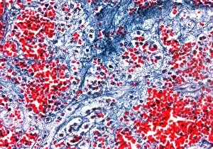
Internal bleeding in lung tissue due to pulmonary emphysema, light micrograph. The bleeding is seen as numerous red blood cells (red)

Background imageRed Blood Cells Collection: Lymph node, SEM

Lymph node, SEM
Lymph node. Coloured scanning electron micrograph (SEM) of a freeze fracture section through a lymph node, showing part of the medullary sinus

Background imageRed Blood Cells Collection: Coloured SEM of a blood clot due to an injury

Coloured SEM of a blood clot due to an injury

Background imageRed Blood Cells Collection: Computer artwork of a blood clot

Computer artwork of a blood clot
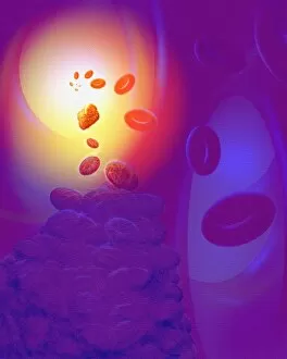
Blood clot. Computer artwork of a blood clot, showing disc-like red blood cells and fibrin. Fibrin (yellow fibres) is a long insoluble protein produced from the blood protein fibrinogen

Background imageRed Blood Cells Collection: Red blood cells embedded in tissue

Red blood cells embedded in tissue
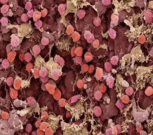
Red blood cells. Coloured scanning electron micrograph of red blood cells in tissue. Blood is composed mainly of red blood cells (erythrocytes). Red blood cells are pumped to all parts of the body

Background imageRed Blood Cells Collection: Blood cells, computer artwork

Blood cells, computer artwork
Blood cells. Computer artwork of red blood cells, white blood, white blood cells (blue) and platelets (yellow) travelling through the lumen of the blood vessel

Background imageRed Blood Cells Collection: Human blood cells, light micrograph

Human blood cells, light micrograph
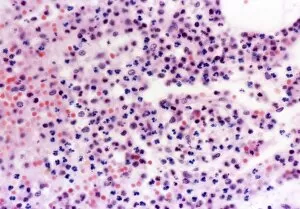
Human blood cells. Light micrograph of blood cells at a site of inflammation, showing red blood cells (erythrocytes, red) surrounded by many granulocytes (blue)


For sale as Licensed Images

Choose your image, Select your licence and Download the media

"Exploring the Intricate World of Red Blood Cells: From Menstruation to Malaria" Uterus lining during menstruation, SEM: Witness the fascinating transformation of the uterus lining during menstruation as red blood cells play a crucial role in this natural process. Red and white blood cells, SEM: Delve into the microscopic realm where red and white blood cells coexist, working together to maintain our body's health and vitality. Blood clot, SEM C016 / 9747: Uncover the intricate structure of a blood clot under scanning electron microscopy, highlighting how red blood cells contribute to its formation. Red blood cells, SEM: Marvel at the mesmerizing beauty of individual red blood cells captured through scanning electron microscopy, showcasing their unique shape and function. Sleeping sickness parasite: Explore how red blood cells become unwitting hosts for parasites causing sleeping sickness, shedding light on this devastating disease's impact on human health. Mouse malaria parasite, SEM: Dive into the world of malaria as we examine mouse malaria parasites residing within red blood cells using scanning electron microscopy techniques. Malaria parasites, TEM: Peer deep inside infected red blood cells with transmission electron microscopy to unravel the complex life cycle and pathogenicity of malaria parasites. Artery SEM: Take a closer look at an artery's inner wall under scanning electron microscopy to understand how healthy red blood cell flow is vital for cardiovascular well-being. Deep vein thrombus: Discover how disrupted circulation can lead to dangerous deep vein thrombosis as clots form within veins due to abnormal interactions between platelets and red blood cells. Histopathology and pathophysiology of diabetic foot ulcers: Gain insights into diabetes-related complications by examining histopathological changes in foot ulcers caused by impaired microcirculation involving damaged capillaries and compromised oxygen supply from reduced numbers or functionality of red blood cells.