
Reproductive Organ Collection
"Exploring the Intricacies of Reproductive Organs: From Spider Lily Stamen to Biomedical Illustrations" Delicate and intricate


For sale as Licensed Images
Choose your image, Select your licence and Download the media
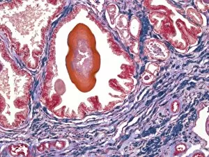
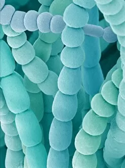
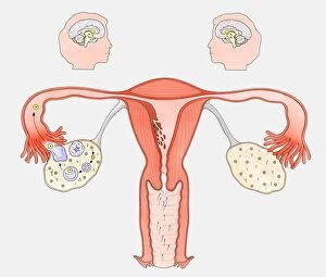
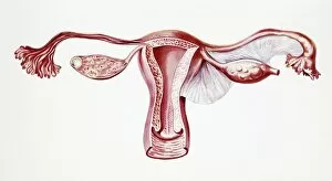
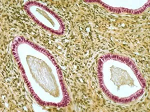
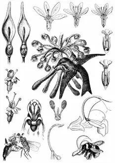
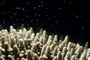
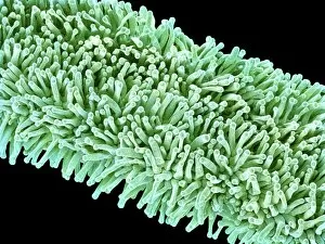
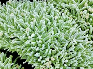
"Exploring the Intricacies of Reproductive Organs: From Spider Lily Stamen to Biomedical Illustrations" Delicate and intricate, the spider lily flower stamen reveals its reproductive prowess under the scanning electron microscope (SEM). Dive into the depths of understanding with a cross-section biomedical illustration showcasing the complexity of the female reproductive system. Euphorbia flower's SEM image unveils its fascinating reproductive parts, offering an up-close look at nature's marvels. Journey inside and witness an interior view of our remarkable reproductive organs, a testament to life's creation. A man wearing undergarments reminds us that male reproductive health is equally important in maintaining overall well-being. Explore how female sexual organs interact with the brain through a comprehensive diagram depicting both normal reproductive cycles and contraceptive pill effects. Light micrograph F006/9805 captures the beauty and intricacy of the cervix, highlighting its vital role in reproduction. Marvel at light micrograph F006/9799 as it unravels the wonders of fallopian tubes, crucial pathways for fertilization to occur. Discover detailed illustrations presenting both male and female reproductive systems, unraveling their unique functions in creating life. A black and white illustration showcases a mature Fucus vesiculosus (Bladderwrack) with swollen tips containing intriguing reproductive organs—nature's secret to pollination revealed. In this captivating journey through various hints about "reproductive organ, " we explore microscopic wonders, biomedical illustrations, human anatomy diagrams, and even delve into nature's own mechanisms for pollination—all reminding us of life's incredible diversity and beauty intertwined within these essential structures.