
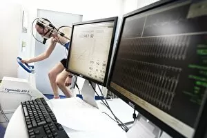
Respiratory Collection (#2)
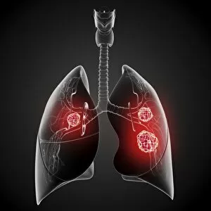
"The Marvels of the Respiratory System: Exploring the Gateway to Life" Diagram of the Lungs and Bronchial Tubes


For sale as Licensed Images
Choose your image, Select your licence and Download the media
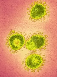
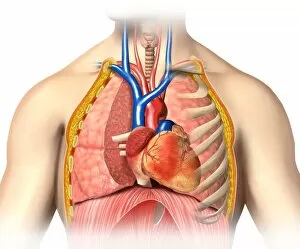
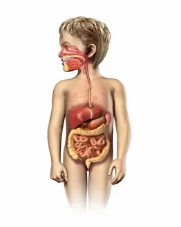
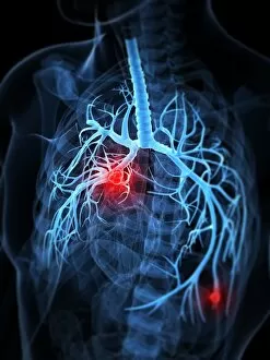
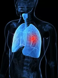
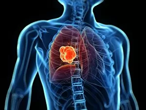
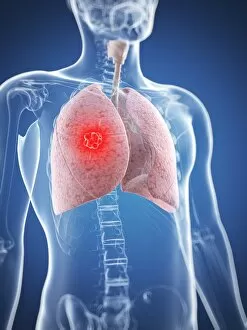
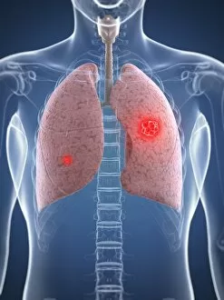
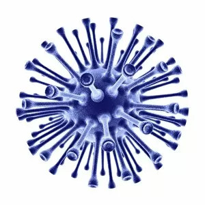
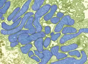
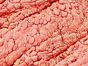
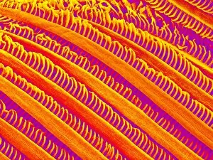
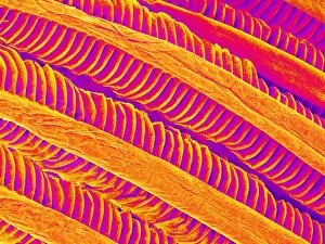
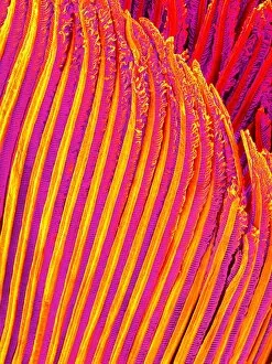
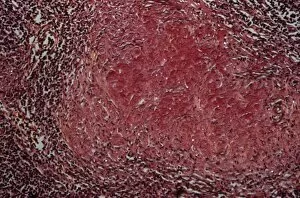
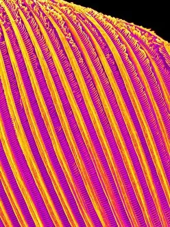
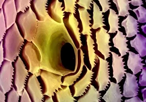
"The Marvels of the Respiratory System: Exploring the Gateway to Life" Diagram of the Lungs and Bronchial Tubes: Unveiling the intricate network within our bodies that facilitates every breath we take, this diagram showcases the lungs and bronchial tubes, highlighting their vital role in oxygenating our bloodstream. Diagram of the Heart, Lungs, and Windpipe: Witnessing a symphony of life-sustaining organs working harmoniously together, this diagram depicts the heart pumping blood while being surrounded by the lungs and windpipe – a testament to our body's remarkable respiratory system. Illustration of Human Respiratory System: Embark on an anatomical journey through our respiratory system as this illustration reveals its various components such as oral cavity, nasal cavity, larynx, trachea, bronchus, and lungs – each playing a crucial part in ensuring efficient respiration. Tension Pneumothorax X-ray: Peering into medical imaging technology's window into diagnosis and treatment strategies for collapsed lungs caused by tension pneumothorax - an urgent condition requiring immediate attention to restore normal breathing patterns. English Oak Leaf Pores SEM Image: Delving into nature's wonders at microscopic levels reveals mesmerizing images showcasing English oak leaf pores under scanning electron microscopy (SEM), reminding us how even plants have their unique respiratory systems. Cystic Fibrosis: Unmasking a Genetic Challenge Exploring cystic fibrosis - a genetic disorder affecting lung function - sheds light on ongoing research efforts aimed at improving treatments for individuals facing challenges with their respiratory health from an early age. Konstantin Buteyko: Soviet Doctor Revolutionizing Breathing Techniques Discovering how Dr. Konstantin Buteyko revolutionized breathing techniques offers insights into his innovative methods that have helped countless individuals manage asthma symptoms and enhance overall respiratory well-being. Paramyxovirus Particles TEM Image.