mail_outline sales@mediastorehouse.com
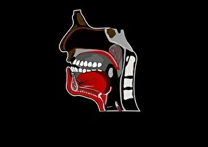
Mouth anatomy, artworkMouth anatomy. Artwork of a section through the human head showing the anatomy of the mouth, nose and upper throat. Above the mouth is the nasal cavity (black)
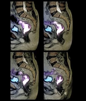
Rectal cancer, MRI scans C018 / 0510Rectal cancer. Coloured magnetic resonance imaging (MRI) scans of the pelvis of a 65 year old patient with a malignant (cancerous) tumour in the rectum, the last part of the large intestine
Knee injury, 3D CT scan C018 / 0648Knee injury. Coloured 3D computed tomography (CT) scan of a knee with a meniscal tear (white line at right between bones)
Knee injury, 3D CT scan C018 / 0639Knee injury. Coloured 3D computed tomography (CT) scan of a knee with a meniscal tear (indicated by arrows). The menisci are fibrocartilage crescents that act as shock absorbers between the femur
Rectal cancer, MRI scans C018 / 0511Rectal cancer. Coloured magnetic resonance imaging (MRI) scans of the pelvis of a 65 year old patient with a malignant (cancerous) tumour (purple) in the rectum, the last part of the large intestine
Knee injury, 3D CT scan C018 / 0645Knee injury. Coloured 3D computed tomography (CT) scan of a knee with a meniscal tear (blue line at right between bones). The menisci are fibrocartilage crescents that act as shock absorbers between
Rectal cancer, MRI scan C018 / 0512Rectal cancer. Coloured magnetic resonance imaging (MRI) scan of the pelvis of a 65 year old patient with a malignant (cancerous) tumour in the rectum, the last part of the large intestine
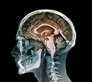
Human head, MRI and 3D CT scans C018 / 0636Human head. Coloured composite image of a magnetic resonance imaging (MRI) scan of the brain and 2D and 3D computed tomography (CT) scans of the head and neck of a 35 year old patient
Anatomical planes of the body, artwork. The three planes shown here are the axial, saggital and coronal. The axial, or transverse, plane splits the body along a horizontal axis
Human spine, artworkHuman spine. Artwork of a healthy human spine. The spine is a column of 33 cylindrical bones, called vertebrae, that support the trunk and head and protect the spinal cord
Choose your image, Select your licence and Download the media