Scanner Collection (#5)

"Unveiling the Hidden Wonders: The Scanner's Journey" Delicate and ethereal

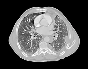
Background imageScanner Collection: Mesothelioma lung cancer, CT scan

Mesothelioma lung cancer, CT scan
Mesothelioma lung cancer. Computed tomography (CT) scan of the lungs of a 69 year old patient with mesothelioma, a type of lung cancer

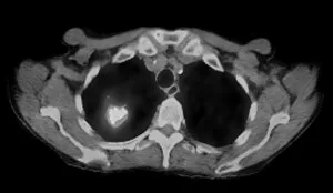
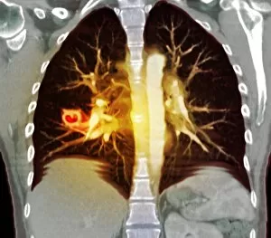
Background imageScanner Collection: Lung cancer, CT and PET scans

Lung cancer, CT and PET scans
Lung cancer. Combined computed tomography (CT) and positron emission tomography (PET) scans of the lungs of an 84 year old woman with a malignant (cancerous) tumour (bright patch)

Background imageScanner Collection: Polycystic kidneys, ultrasound scan C017 / 7747

Polycystic kidneys, ultrasound scan C017 / 7747
Polycystic kidneys. Ultrasound scan of the kidneys of a 42 year old female showing numerous fluid-filled cysts (dark areas)

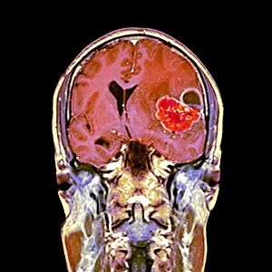
Background imageScanner Collection: Brain cancer, MRI scan C016 / 4438

Brain cancer, MRI scan C016 / 4438
Brain cancer. Coloured coronal magnetic resonance imaging (MRI) scan through the head of a 48 year old male patient with a glioblastoma (red)

Background imageScanner Collection: Brain cancer, MRI scan C016 / 4436

Brain cancer, MRI scan C016 / 4436
Brain cancer. Coloured coronal magnetic resonance imaging (MRI) scan through the head of a 48 year old male patient with a glioblastoma (centre right)

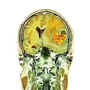
Background imageScanner Collection: Brain cancer, MRI scan C016 / 4437

Brain cancer, MRI scan C016 / 4437
Brain cancer. Coloured coronal magnetic resonance imaging (MRI) scan through the head of a 48 year old male patient with a glioblastoma (orange)

Background imageScanner Collection: QR code after being scanned C013 / 9472

QR code after being scanned C013 / 9472
QR code (on bottle) after being scanned by a mobile phone. A QR code, designed to be scanned by devices such as mobile phones
Background imageScanner Collection: QR code being scanned C013 / 9473

QR code being scanned C013 / 9473
QR code (lower right) being scanned by a tablet computer. A QR code, designed to be scanned by devices such as mobile phones or mobile-enabled computers
Background imageScanner Collection: QR code being scanned C013 / 9471

QR code being scanned C013 / 9471
QR code (on bottle) being scanned by a mobile phone. A QR code, designed to be scanned by devices such as mobile phones, is a matrix barcode that consists of black modules arranged in a square

Background imageScanner Collection: Storm chasing, Texas, USA

Storm chasing, Texas, USA
Storm chasing. Tornado researcher monitoring a severe thunderstorm from inside a Doppler on Wheels (DOW) portable weather radar truck

Background imageScanner Collection: Storm chasing, USA

Storm chasing, USA
Storm chasing. Close-up of the undercarriage of a Doppler on Wheels (DOW) portable weather radar truck, during a severe storm

Background imageScanner Collection: Storm chasing, South Dakota, USA

Storm chasing, South Dakota, USA
Storm chasing. Doppler on Wheels (DOW) portable weather radar truck scanning a severe thunderstorm. This truck is one of more than 100 research vehicles that participated in VORTEX 2

Background imageScanner Collection: Storm chasing, Nebraska, USA

Storm chasing, Nebraska, USA
Storm chasing. Doppler on Wheels (DOW) portable weather radar truck scanning a severe thunderstorm. This truck is one of more than 100 research vehicles that participated in VORTEX 2

Background imageScanner Collection: Biometric DNA scan, computer artwork

Biometric DNA scan, computer artwork
Biometric DNA scanner. Computer artwork of a DNA scanner being used to verify a persons identity. Security technology based on recognising an individuals biological data, such as fingerprints, iris

Background imageScanner Collection: Fingerprint scanner, computer artwork

Fingerprint scanner, computer artwork
Fingerprint scanner. Computer artwork of a scanner confirming the identity of a persons fingerprint. Security technology based on identifying a persons unique biological data such as fingerprints

Background imageScanner Collection: Thyroid gland, scintigram

Thyroid gland, scintigram
Thyroid gland. Coloured scintigram of a healthy thyroid gland. This two-lobed endocrine gland is found in the neck, where it lies on top of the trachea (windpipe)

Background imageScanner Collection: Healthy kidneys, CT scan

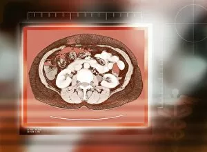
Healthy kidneys, CT scan
Healthy kidneys, coloured computed tomography (CT) scan. The kidneys are the two C-shaped white organs either side of the spinal vertebra (centre). Below the spine and kidneys are back muscles

Background imageScanner Collection: Knee joint, side view, MRI scan

Knee joint, side view, MRI scan
Knee joint, coloured magnetic resonance imaging (MRI) scan. In this side view, the front of the knee is at left. The patella (kneecap) is at far left. The knee is the largest joint in the body

Background imageScanner Collection: Female pelvis, coloured 3-D CT scan

Female pelvis, coloured 3-D CT scan
Female pelvis, coloured 3-D computed tomography (CT) scan. The upper part of each thigh bone (femur) is seen at each side of the pelvis, where they form the hip joints

Background imageScanner Collection: Abdominal bones, 3-D CT scan

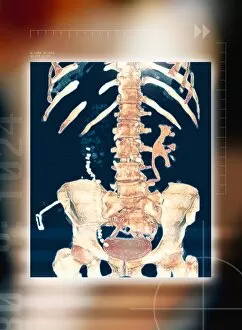
Abdominal bones, 3-D CT scan
Abdominal bones, coloured 3-D computer tomography (CT) scan. The lower part of the ribcage is in upper frame, with the pelvis in lower frame. They are connected by the spine, which runs down centre

Background imageScanner Collection: Brain and spinal cord, MRI

Brain and spinal cord, MRI
Brain and spinal cord. Coloured magnetic resonance imaging (MRI) scan of a healthy brain (blue) and upper spinal cord (centre to lower centre)

Background imageScanner Collection: Artwork of home computer & collage of peripherals

Artwork of home computer & collage of peripherals
Home computer & peripherals. Artwork showing a collage of computer peripherals around a home computer. The computer (at centre) has a mock roof to signify a home computer

Background imageScanner Collection: Ultrasound transducer

Ultrasound transducer
MODEL RELEASED. Ultrasound transducer. This device emits ultrasonic waves and detects the reflected echoes. The change in frequency is used to build images of the inside of the body

Background imageScanner Collection: Doppler ultrasound scanning of carotid artery

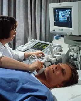
Doppler ultrasound scanning of carotid artery

Background imageScanner Collection: Ultrasound scanning of neck blood vessels

Ultrasound scanning of neck blood vessels

Background imageScanner Collection: Early television system, 19th century

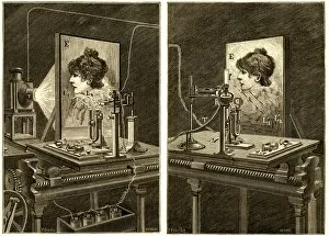
Early television system, 19th century
Early television system. Historical artwork of a proposed 19th century system to transmit an image by using optical and telephone technology. The original image (a womans head) is at left

Background imageScanner Collection: Brain scanning, conceptual artwork

Brain scanning, conceptual artwork. This may represent the brain being scanned by a CT (computed tomography) or MRI (magnetic resonance imaging) scanner

Background imageScanner Collection: Ultrasound scanner and scans, artwork

Ultrasound scanner and scans, artwork
Ultrasound scanning, conceptual artwork. The scanner itself is at centre. Ultrasound scanning uses high frequency sound waves to image the body

Background imageScanner Collection: Podiatric ultrasound

Podiatric ultrasound
MODEL RELEASED. Podiatric ultrasound. Podiatrist performing an ultrasound examination on the sole of a foot. This technique is used to diagnose soft tissue problems in the foot

Background imageScanner Collection: Artwork of a hand holding an ultrasound transducer

Artwork of a hand holding an ultrasound transducer
Ultrasound transducer. Computer artwork of a hand holding an ultrasound transducer. The transducer can be used as a diagnostic tool for foetal scanning

Background imageScanner Collection: Doppler ultrasound scanning of a mans abdomen

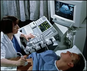
Doppler ultrasound scanning of a mans abdomen
MODEL RELEASED. Ultrasound scanning. Doctor uses a doppler ultrasound scanner to examine a blood vessel in a mans abdomen

Background imageScanner Collection: Fractured atlas vertebra, 3D CT scan

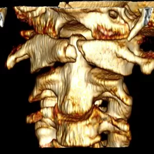
Fractured atlas vertebra, 3D CT scan
Fractured atlas vertebra, coloured 3D CT (computed tomography) scan. Posterior view of a fractured atlas vertebra (break at upper centre) of a 34 year old man. Part of the skull can be seen at top

Background imageScanner Collection: Knee meniscus tear

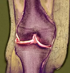
Knee meniscus tear
MODEL RELEASED. Knee meniscus tear. Coloured magnetic resonance imaging (MRI) scan of a coronal (front) section through the right knee joint of a 52 year old woman

Background imageScanner Collection: Pancreatitis, CT scan

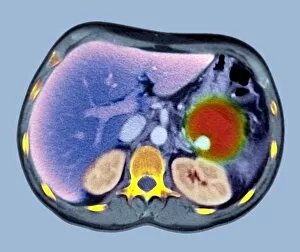
Pancreatitis, CT scan
Pancreatitis. Coloured computed tomography (CT) scan of an axial section through a patients abdomen showing a false aneurysm (red) due to chronic pancreatitis. The front of the body is at top

Background imageScanner Collection: Lung lesions, CT scan

Lung lesions, CT scan
Lung lesion. Coloured frontal computed tomography (CT) scan of a woman with Wegeners granulomatosis, showing cavitating (cavity forming) lesions (left, orange) in the lungs (black)

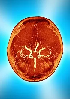
Background imageScanner Collection: Brain and arteries, coloured CT scan

Brain and arteries, coloured CT scan
Healthy brain and arteries. Coloured computed tomography (CT) scan showing healthy arteries (white) supplying a human brain (red) with oxygenated blood. The front of the head is at top

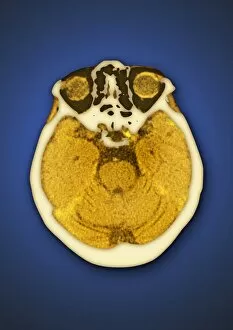
Background imageScanner Collection: Healthy brain and frontal sinus, CT scan

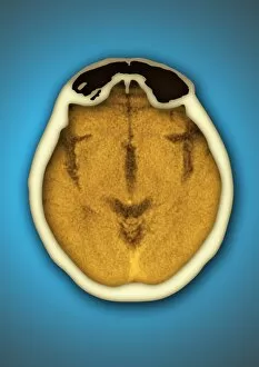
Healthy brain and frontal sinus, CT scan
Healthy brain and frontal sinus. Coloured computed tomography (CT) scan of a human head. The axial (horizontal) scan has revealed the head and brain in cross section. The front of the head is at top

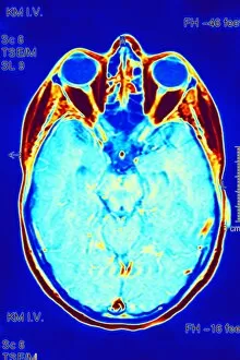
Background imageScanner Collection: Healthy brain and eyes, CT scan

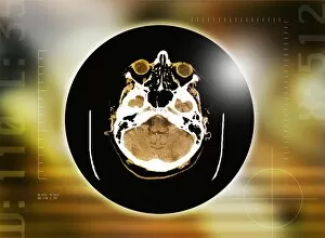
Healthy brain and eyes, CT scan
Healthy brain and eyes. Coloured computed tomography (CT) scan of a human head. The axial (horizontal) scan has revealed the head and brain in cross section at eye-level

Background imageScanner Collection: Brain scan, MRI scan

Brain scan, MRI scan
Brain scan. Coloured magnetic resonance imaging (MRI) scan of an axial (horizontal) section through a healthy brain. The front of the brain is at top

Background imageScanner Collection: Brain scans, MRI scans

Brain scans, MRI scans
Brain scans. Magnetic resonance imaging (MRI) scans of axial (horizontal) sections through a healthy brain. The front of the brain is towards top left

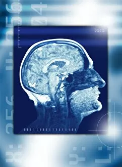
Background imageScanner Collection: Healthy brain, MRI scan

Healthy brain, MRI scan
Healthy brain, coloured magnetic resonance imaging (MRI) scan. The brain is seen in profile. The nose is at far right, and behind it are the sinuses (dark), air spaces in the skull

Background imageScanner Collection: Healthy brain, CT scan

Healthy brain, CT scan
Healthy brain, coloured computed tomography (CT) scan. The front of the brain is at top. The brown circles are the eyeballs

Background imageScanner Collection: Brain and blood vessels, 3-D CT scan

Brain and blood vessels, 3-D CT scan
Brain and blood vessels, coloured 3-D computed tomography (CT) scan. The front of the brain is at bottom. It is enclosed by the skull (grey/green). Blood vessels (red) supply the brain with blood

Background imageScanner Collection: Head, side view, MRI scan

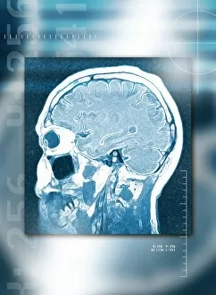
Head, side view, MRI scan
Head from the side, magnetic resonance imaging (MRI) scan. The brain (light blue, folded) fills the cerebrum of the skull (white). At centre left is an eyeball (dark blue circle)

Background imageScanner Collection: Skull, 3-D CT scan

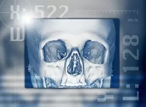
Skull, 3-D CT scan
Human skull, 3-D computed tomography (CT) scan. The orbits of the eyes are either side of the nasal cavity. The front of the skull contains large air spaces called sinuses

Background imageScanner Collection: Head, CT scans

Head, CT scans
Head scans. Coloured computed tomography (CT) scans of axial sections through a healthy head. The sequence of scans (from left to right)

Background imageScanner Collection: MRI scanner

MRI scanner. Computer artwork of an MRI (magnetic resonance imaging) scanner with a female patient. The patient is passed inside the machine (through the hole, upper left) to be scanned

Background imageScanner Collection: High resolution X-ray CT scanner

High resolution X-ray CT scanner
High resolution X-ray computed tomography (CT) scanner, University of Texas, Austin, USA. This scanner is used by scientists studying rocks, fossils and biological specimens


For sale as Licensed Images

Choose your image, Select your licence and Download the media

"Unveiling the Hidden Wonders: The Scanner's Journey" Delicate and ethereal, a sea green swallowtail butterfly gracefully flutters its wings under the watchful eyes of a scanner. With precision and accuracy, a full body scan captures every detail, revealing the intricate workings of our complex human form. Peering into the depths of brain anatomy, an MRI scan unveils the mysteries hidden within, unlocking secrets that shape our thoughts and actions. Like a unique signature, a fingerprint scanner maps out individual patterns with meticulous care, ensuring security in an ever-evolving world. Injured but not defeated, an MRI C018 / 0649 reveals the extent of damage to a ruptured Achilles tendon - guiding doctors towards effective treatment options. Beyond fingerprints' utilitarian purpose lies artistic beauty; an artwork created by capturing each swirl through a fingerprint scanner tells tales untold before. A glimpse into normalcy is found as an MRI C016 / 8845 showcases the intricacies of a healthy human brain - reminding us of its remarkable resilience. Seeking answers beneath layers unseen, an MRI exposes ruptured breast implants - providing clarity for those seeking resolution and healing. Innocence encapsulated within imaging technology as we witness childlike wonder through glimpses into young minds via captivating MRI scans. Unraveling connections that make us who we are; white matter fibers come alive under DTI scans - illuminating pathways crucial for communication within our brains. Echoing familiarity amidst diversity; another snapshot from everyday life emerges with Normal Human Brain captured by MRI C016 / 8850 – reminding us how extraordinary ordinary can be.