Scanning Electron Microscope Collection (#97)

"Unveiling the Hidden World

Background imageScanning Electron Microscope Collection: Plant pores, SEM

Plant pores, SEM
Plant pores. Coloured scanning electron micrograph (SEM) of stomata (pores) on the pistil of a bellflower (Adenophora sp.). Stomata are the site of gaseous exchange, or respiration

Background imageScanning Electron Microscope Collection: Germinating red chard seed, SEM

Germinating red chard seed, SEM
Germinating red chard seed. Coloured scanning electron micrograph (SEM) of a red chard seed (Beta vulgaris var. cicla) sprouting a root (white)

Background imageScanning Electron Microscope Collection: Primrose pollen, SEM

Primrose pollen, SEM
Primrose pollen. Coloured scanning electron micrograph (SEM) of pollen grains (pink) from a primrose flower (Primula vulgaris)

Background imageScanning Electron Microscope Collection: Open stoma, SEM

Open stoma, SEM
Open stoma. Coloured scanning electron micrograph (SEM) of an open stoma on the surface of a tobacco leaf (Nicotiana tabacum)

Background imageScanning Electron Microscope Collection: Cryptotaenia japonica Hassk flowers, SEM

Cryptotaenia japonica Hassk flowers, SEM
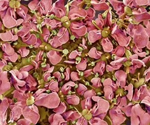
Cryptotaenia japonica Hassk flowers. Coloured scanning electron micrograph (SEM) of the flowers of Japanese hornwort (Cryptotaenia japonica Hassk)

Background imageScanning Electron Microscope Collection: Bacteria in the nose, SEM

Bacteria in the nose, SEM
Bacteria in the nose. Coloured scanning electron micrograph (SEM) of bacteria (red) on the surface of the nasal cavity. One of the squamous epithelium cells (lower centre to lower right)

Background imageScanning Electron Microscope Collection: Tongue bacteria, SEM

Tongue bacteria, SEM
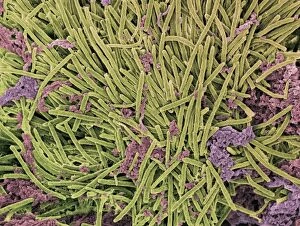
Tongue bacteria. Coloured scanning electron micrograph (SEM) of bacteria on the surface of a human tongue. Large numbers of bacteria can form a visible layer on the surface of the tongue

Background imageScanning Electron Microscope Collection: Vibrio vulnificus bacteria, SEM

Vibrio vulnificus bacteria, SEM
Vibrio vulnificus bacteria, coloured scanning electron micrograph (SEM). This Gram-negative rod- shaped motile bacteria is found in sea water

Background imageScanning Electron Microscope Collection: Proteus vulgaris bacteria, SEM

Proteus vulgaris bacteria, SEM
Proteus vulgaris bacteria, scanning electron micrograph (SEM). The fine threadlike projections are flagella, which allow the bacterium to move. P

Background imageScanning Electron Microscope Collection: Gonorrhoea bacteria, SEM

Gonorrhoea bacteria, SEM
Gonorrhoea bacteria. Coloured scanning electron micrograph (SEM) of gonorrhoea bacteria (Neisseria gonorrhoeae, round) on a human epithelial cell

Background imageScanning Electron Microscope Collection: Tobacco plant protoplast, SEM

Tobacco plant protoplast, SEM
Protoplast. Coloured scanning electron micrograph (SEM) of a tobacco plant (Nicotiana tabacum) protoplast. A protoplast is a plant cell that has had its tough outer cell wall removed by chemical

Background imageScanning Electron Microscope Collection: Chinese lantern flower skeleton, SEM

Chinese lantern flower skeleton, SEM
Chinese lantern flower skeleton. Coloured scanning electron micrograph (SEM) of the decaying calyx of a flower of the chinese lantern plant (Physalis sp.)

Background imageScanning Electron Microscope Collection: Uranyl acetate crystals, SEM

Uranyl acetate crystals, SEM
Uranyl acetate. Coloured scanning electron micrograph (SEM) of rhombic crystals of uranyl acetate. This chemical, made from depleted uranium, is used as a negative stain in electron microscopy

Background imageScanning Electron Microscope Collection: Salt grain, SEM

Salt grain, SEM
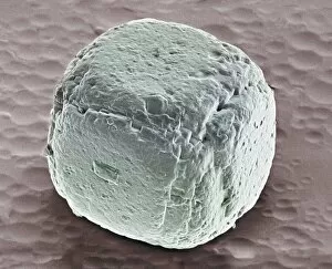
Salt grain, coloured scanning electron micrograph (SEM). A salt (sodium chloride) crystal is composed of a cubic lattice of sodium and chloride ions. Salt is commonly used as seasoning in cooking

Background imageScanning Electron Microscope Collection: Cell infected with HIV, SEM

Cell infected with HIV, SEM
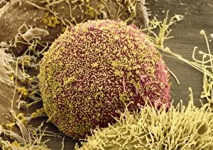
Cell infected with HIV. Coloured scanning electron micrograph (SEM) of HIV particles (yellow) budding from the membrane of a host cell (red)

Background imageScanning Electron Microscope Collection: Salt grains, SEM

Salt grains, SEM
Salt grains, coloured scanning electron micrograph (SEM). Each salt (sodium chloride) crystal is composed of a cubic lattice of sodium and chloride ions. Salt is commonly used as seasoning in cooking

Background imageScanning Electron Microscope Collection: Maggot skin, SEM

Maggot skin, SEM
Maggot skin, coloured scanning electron micrograph (SEM). Maggots are the legless larvae of flies (order Diptera). Here, the skin of the maggot is covered in fine hooks which enables it to grip a

Background imageScanning Electron Microscope Collection: Chaetognath, SEM

Chaetognath, SEM
Chaetognath. Scanning electron micrograph (SEM) of a chaetognath (phylum Chaetognatha). This microscopic predatory worm is transparent or translucent and is covered by a cuticle

Background imageScanning Electron Microscope Collection: Wasp foot, SEM

Wasp foot, SEM
Wasp foot. Coloured scanning electron micrograph (SEM) of the foot of a common wasp (Vespula vulgaris). This is the final segment of the leg, known as the tarsus

Background imageScanning Electron Microscope Collection: Compound eye of a gnat, SEM

Compound eye of a gnat, SEM
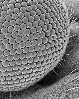
Compound eye of a gnat. Scanning electron micrograph (SEM) of the surface of a compound eye from a fungus gnat (suborder Nematocera). The eye consists of many rounded lenses known as ommatidia

Background imageScanning Electron Microscope Collection: Head of a pipistrelle bat

Head of a pipistrelle bat. Coloured scanning electron micrograph (SEM) of the head of a pipistrelle bat (Pipistrellus pipistrellus)

Background imageScanning Electron Microscope Collection: Bat tick mouthparts, SEM

Bat tick mouthparts, SEM
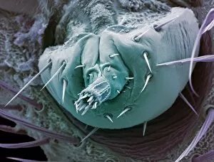
Bat tick mouthparts. Coloured scanning electron micrograph (SEM) of a bat tick (order Acarina), an arachnid ectoparasite (external parasites) that feeds on the blood of bats

Background imageScanning Electron Microscope Collection: Woven silk, SEM

Woven silk, SEM
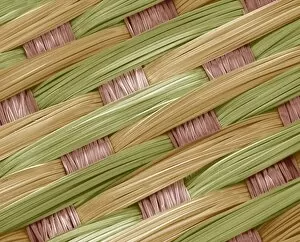
Woven silk, coloured scanning electron micrograph (SEM). Silk is a protein fibre, which can be woven into textiles. It is obtained from the cocoons of metamorphosising silkworms

Background imageScanning Electron Microscope Collection: Aspirin crystals, SEM

Aspirin crystals, SEM
Aspirin crystals, coloured scanning electron micrograph (SEM). Aspirin (acetylsalicylic acid) is a non-steroidal anti-inflammatory drug (NSAID)

Background imageScanning Electron Microscope Collection: Antihistamine drug crystals, SEM

Antihistamine drug crystals, SEM
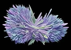
Antihistamine drug crystals, coloured scanning electron micrograph (SEM). Antihistamines block the effect of the chemical histamine

Background imageScanning Electron Microscope Collection: Mescaline crystals, SEM

Mescaline crystals, SEM
Mescaline. Coloured scanning electron micrograph (SEM) of crystals of mescaline. Mescaline is an illegal hallucinogenic drug produced from the dried tops, or buttons

Background imageScanning Electron Microscope Collection: Salbutamol sulphate crystals, SEM

Salbutamol sulphate crystals, SEM
Salbutamol sulphate crystals, coloured scanning electron microscope (SEM). Salbutamol sulphate (ventolin) is a bronchodilator drug used to treat asthma

Background imageScanning Electron Microscope Collection: Bronchodilator drug crystals, SEM

Bronchodilator drug crystals, SEM
Bronchodilator drug crystals, coloured scanning electron micrograph (SEM). These are crystals of the drug ventolin (salbutamol sulphate), a bronchodilator drug used to treat asthma

Background imageScanning Electron Microscope Collection: Sea salt, SEM

Sea salt, SEM
Sea salt. Coloured scanning electron micrograph (SEM) of sea salt, showing its crystalline structure. Sea salt consists mainly of sodium chloride (NaCl), but unlike pure table salt

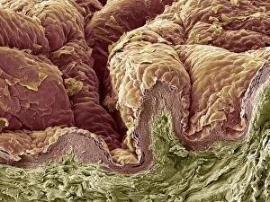
Background imageScanning Electron Microscope Collection: Skin tissue, SEM

Skin tissue, SEM
Skin tissue. Coloured scanning electron micrograph (SEM) of a freeze-fracture through human skin tissue. The fracture plane (lower frame) has revealed the pseudo-stratified epithelium (below surface)

Background imageScanning Electron Microscope Collection: Nib of a ballpoint pen, SEM

Nib of a ballpoint pen, SEM
Nib of a ballpoint pen, coloured scanning electron micrograph (SEM). Ballpoint pens have an internal chamber filled with a viscous ink that is dispensed at the tip during use by the rolling action of

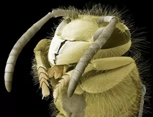
Background imageScanning Electron Microscope Collection: Common wasp head, SEM

Common wasp head, SEM
Common wasp (Vespula vulgaris) head, coloured scanning electron micrograph (SEM). The antennae are seen on the top of the head, between the two large compound eyes

Background imageScanning Electron Microscope Collection: Wasp tongue surface, SEM

Wasp tongue surface, SEM
Wasp tongue surface. Coloured scanning electron micrograph (SEM) of the surface of the tongue of a wasp. Unlike the equivalent structure in bees (the proboscis)

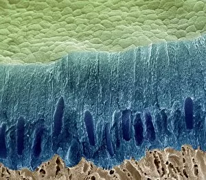
Background imageScanning Electron Microscope Collection: Tooth enamel formation, SEM

Tooth enamel formation, SEM
Tooth enamel formation. Coloured scanning electron micrograph (SEM) of a freeze-fractured section through a tooth, showing the enamel-forming cell layer (blue)


For sale as Licensed Images

Choose your image, Select your licence and Download the media

"Unveiling the Hidden World: Exploring with a Scanning Electron Microscope" The scanning electron microscope (SEM) is an incredible tool that allows us to delve into the microscopic realm, revealing intricate details and unlocking secrets of the unseen. With its high magnification capabilities, it offers a glimpse into fascinating aspects of life and nature. In one captivating image, we witness the beauty of gender determination as X and Y chromosomes are captured under the SEM's lens. The intricate patterns and structures within these chromosomes showcase the complexity of our genetic makeup. Moving on to another astonishing discovery, a Scanning Electron Micrograph presents us with an up-close view of a Praying Mantis at 30 times magnification. Every tiny detail becomes visible, from its delicate limbs to its mesmerizing eyes – reminding us of nature's remarkable diversity. Shifting focus to plant life, lavender pollen grains come alive in another SEM image. These minuscule particles take center stage as their unique shapes and textures become apparent under intense scrutiny. Zooming in even further reveals a Tardigrade or 'Water Bear' at an astounding 1250 times magnification. This resilient creature captures our imagination as we explore its alien-like features through this powerful imaging technique. Delving deeper into biological wonders, T lymphocytes battling cancer cells are showcased in yet another breathtaking SEM capture. This visual representation highlights the ongoing fight within our bodies against diseases like cancer – emphasizing both hope and resilience. Calcareous phytoplankton takes center stage next as we uncover their intricate forms through SEM imagery. These microscopic organisms play crucial roles in marine ecosystems while leaving us awe-inspired by their stunningly detailed structures. Transitioning to neural marvels, nerve cells reveal their intricacy under the watchful eye of an SEM. Their branching extensions create complex networks that enable communication throughout our bodies – showcasing nature's ingenuity at work. Venturing into insect realms once again, a fruit fly is immortalized under the SEM's lens.