
Scans Collection
"Unveiling the Complexities: Exploring Scans from Human Brains to Livestock" Delving into the Depths


For sale as Licensed Images
Choose your image, Select your licence and Download the media
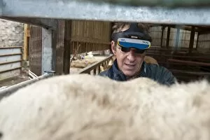
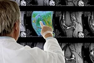
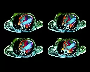
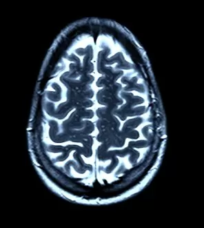
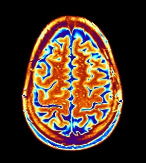
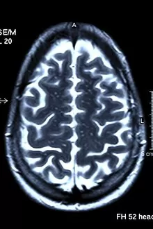
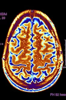
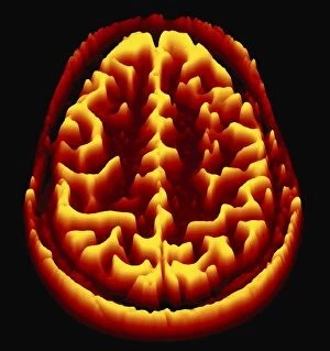
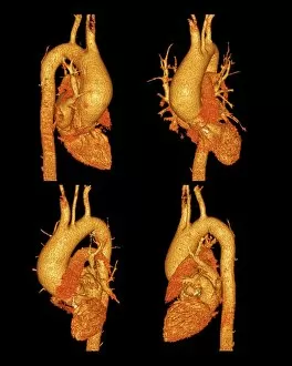
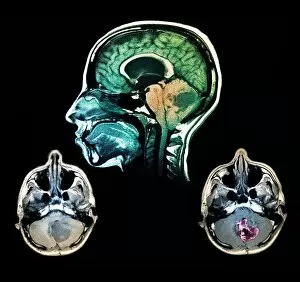
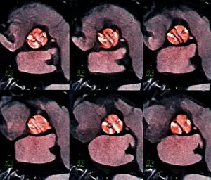
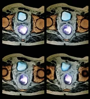
"Unveiling the Complexities: Exploring Scans from Human Brains to Livestock" Delving into the Depths: Unraveling the Mysteries of the Human Brain through 3D-MRI Scans Cutting-Edge Technology in Cattle Farming: Vet Uses Ultrasonic Scanner to Detect Pregnancy, Witnessed Through High-Tech Headset Precision Diagnosis: Radiologist Examines Knee MRI Scans with Expertise and Care Battling Cardiac Lymphoma: A Glimpse into Life-Saving MRI Scans for Heart Health Empathy in Action: Doctor Examining a Patient's Scan with Compassion and Dedication Ensuring Safety and Security: Veterinarian Utilizes Microchip Scan on Domestic Dog at Veterinary Surgery Nurturing New Life on Sheep Farms: Early Stage Pregnancy Revealed by Ultrasound Scanner Screen Counting Blessings, One Lamb at a Time: Ewe's Early Stage Pregnancy Unveiled through Advanced Scanning Techniques Peering Inside Mindscape Masterpieces.