
Secondary Collection (#5)
"Exploring the Spectrum of Knowledge: From Colour Wheels to Cholera Toxins" In this captivating journey through knowledge


For sale as Licensed Images
Choose your image, Select your licence and Download the media
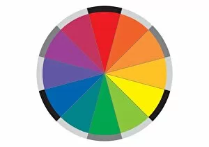
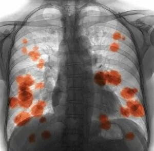
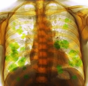
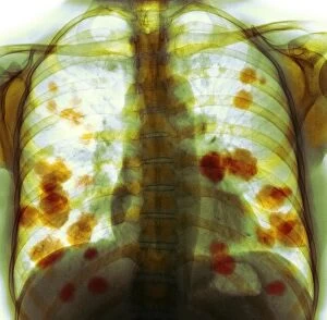
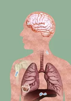
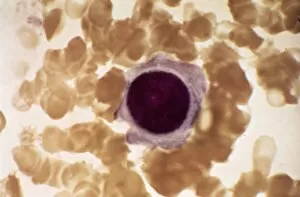
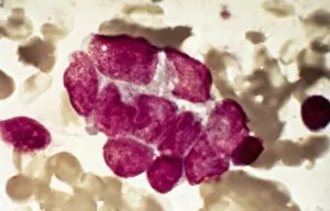
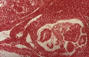
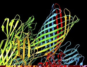
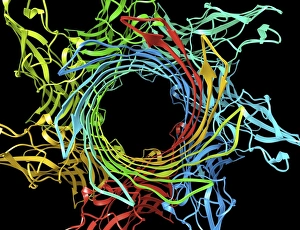
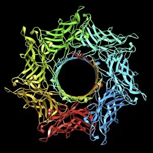
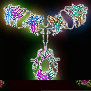
"Exploring the Spectrum of Knowledge: From Colour Wheels to Cholera Toxins" In this captivating journey through knowledge, we delve into the world hints that unveil a tapestry of fascinating subjects. Starting with the vibrant hues of a colour wheel, we are reminded of the intricate interplay between shades and tones that bring life to our visual experiences. Just as Marie Curie's groundbreaking discoveries illuminated the path towards understanding radiation and its effects on humanity. From Montrose Academy in Scotland to Malvern Girls College in Worcestershire, educational institutions have been instrumental in nurturing young minds and shaping future generations. Acland Burghley School in North London witnessed an extraordinary moment on May 24th, 1968 when interviewer Anne Mountfield engaged with youngsters destined for television stardom. Peering through a light micrograph C015/7103 reveals ovarian cancer at a microscopic level, reminding us of the ongoing battle against this devastating disease. Meanwhile, an engraving from Grammar School at Dorchester takes us back to Oxfordshire in 1827, showcasing how education has evolved over centuries. Delving deeper into scientific realms, we encounter molecular models such as cholera toxin and human serum albumin molecule. These intricate structures unlock secrets about diseases and vital bodily functions while paving the way for medical advancements. Finally, a lithographic plate adorned with arms symbolizes heritage and lineage—a reminder that history shapes our present identities. These secondary hints intertwine diverse fields—artistry meets science; academia merges with societal impact—to form a rich tapestry of knowledge waiting to be explored by curious minds seeking enlightenment beyond boundaries.