
Sem Image Collection (#2)
"Exploring the Microcosmos: SEM Image Unveils Nature's Intricate Wonders" Delving into the hidden realms of nature


For sale as Licensed Images
Choose your image, Select your licence and Download the media
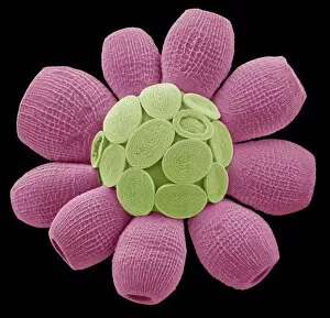
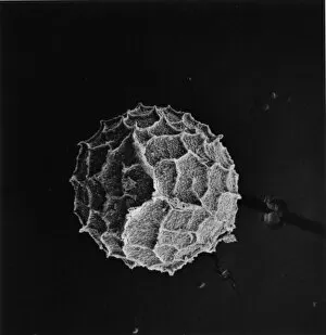
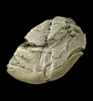
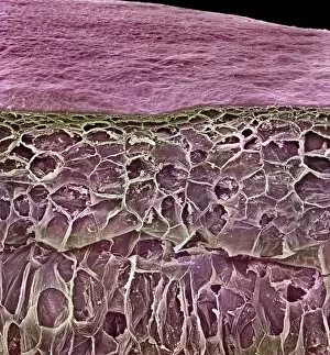
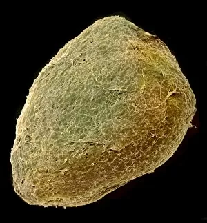
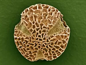
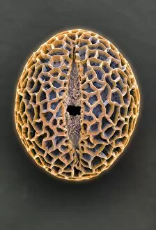
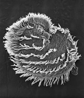
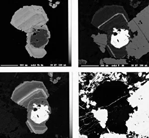
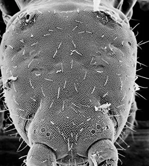
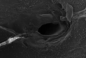
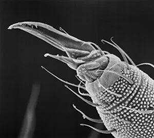
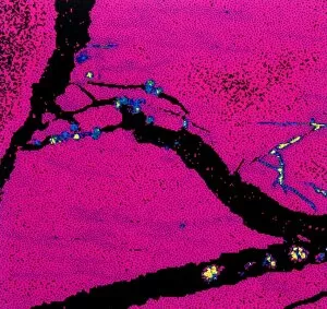
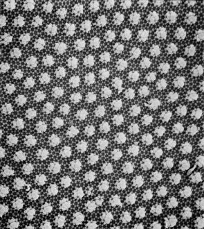
"Exploring the Microcosmos: SEM Image Unveils Nature's Intricate Wonders" Delving into the hidden realms of nature, this captivating SEM image showcases a diverse array of microscopic wonders. Starting with Crysotile asbestos, its delicate fibers resemble an ethereal dance frozen in time. Moving on to the liver, intricate cellular structures come alive under high magnification, revealing the organ's remarkable complexity. The notorious bed bug, Cimex lectularius, appears menacingly detailed as its exoskeleton is meticulously captured by this powerful imaging technique. Meanwhile, Taraxacum officinale's fruiting head exhibits astonishing symmetry and intricacy that belie its humble appearance. Snail teeth are unveiled in all their glory; these minuscule yet robust structures demonstrate nature's ingenuity at creating tools for survival. The Plasmodium sp. , responsible for malaria infections worldwide, reveals itself as a haunting reminder of the ongoing battle against infectious diseases. Kaolinite crystals shimmer like tiny jewels when observed up close – a testament to Earth's geological beauty. Sarcoptes scabiei mites appear almost alien-like with their bristly appendages and segmented bodies. Aspergillus fungi showcase their distinctive branching hyphae network while caterpillar eggs offer a glimpse into new life waiting to emerge. Blackfly antennae stand out with their fine sensory hairs designed for navigation through air currents. Finally, human red blood corpuscles mesmerize with their vibrant hue and unique biconcave shape that enables efficient oxygen transport throughout our bodies. In this extraordinary journey through unseen dimensions, SEM images unveil Mother Nature's exquisite craftsmanship and remind us of her boundless creativity even within the tiniest organisms or structures.