mail_outline sales@mediastorehouse.com
Anatomy of prostate gland
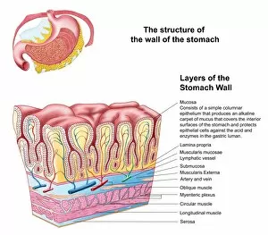
Anatomy of the structure and layers of the stomach wall
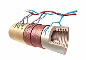
Cross section illustration of human small intestine showing muscle layer, villus and plicae
Cross section illustration of human large intestine
Fallopian tube section, light micrographFallopian tube section. Coloured light micrograph of a section through the ampulla of a fallopian tube. The fallopian tube, or oviduct, conveys the egg from the ovary to the uterus
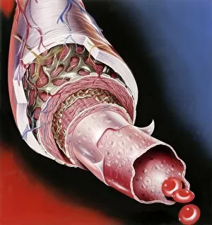
Artery anatomy, artworkArtery anatomy, computer artwork. At the centre of the artery is the lumen, with three red blood cells (erythrocytes, bottom right)
Omentum, light micrograph. The omentum is a fold of peritoneal membrane. Blood vessels are seen criss-crossing the serous membrane. Magnification: x100 when printed at 10 centimetres wide
Intestinal anatomy, artworkIntestinal anatomy. Computer artwork showing the layers of the small intestine. The central space (lumen) is surrounded by the mucosa (beige, folded), which has numerous folds (villi)
Choose your image, Select your licence and Download the media