
Sheath Collection (#5)
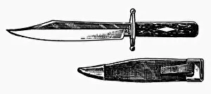
"The Kings' Two Daggers: One with a Blade of Gold, the Other of Iron" In the realm of kings and warriors, two daggers stood as symbols of power


For sale as Licensed Images
Choose your image, Select your licence and Download the media
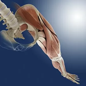
"The Kings' Two Daggers: One with a Blade of Gold, the Other of Iron" In the realm of kings and warriors, two daggers stood as symbols of power. One gleamed with a blade forged from pure gold, radiating opulence and authority. The other possessed an iron edge, representing strength and resilience in battle. These contrasting weapons were not only tools for conquest but also reflections of their wielders' character. Delving deeper into the mysteries of human anatomy, scientists uncovered the intricate process known as myelination. Through Transmission Electron Microscopy (TEM), they captured Picture No. 10876591 - a mesmerizing glimpse into nerve fibers adorned with myelin sheaths that enhance electrical conductivity within our bodies. This microscopic world revealed nature's ingenious design to optimize communication between neurons. Beyond scientific marvels, artists found inspiration in crafting designs for dagger sheaths that transcended mere functionality. A daring creation emerged—a sheath depicting Death itself—an embodiment of mortality juxtaposed against the lethal beauty concealed within. Meanwhile, Regatta and Carnival Week brought joyous festivities to Fowey, Cornwall in August 1993. Amidst vibrant celebrations filled with laughter and merriment, one could almost forget the existence of daggers or their somber purpose. Traveling across borders to Germany's Aachen Cathedral Treasury unveiled Oliphant—a magnificent relic from times long past—its ornate carvings telling tales lost to history while guarding secrets untold. Fashion too had its rendezvous with daggers; Marcus dress advertised Courtelle fabric through an elegant portrayal where sophistication met utility—the perfect fusion for modern women seeking both style and convenience. But let us journey back further still—to medieval England under King Henry III's reign—where three soldiers donned armor bearing witness to countless battles fought on blood-soaked fields; their trusty blades safely nestled within sturdy sheaths at their sides.