
Spermatozoa Collection (#2)
"Spermatozoa: The Tiny Warriors of Life" In the vast world of reproductive biology, spermatozoa play a pivotal role in the creation of life


For sale as Licensed Images
Choose your image, Select your licence and Download the media
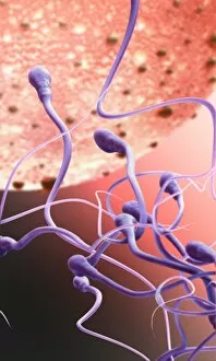
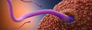
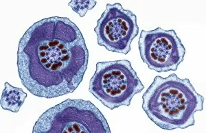
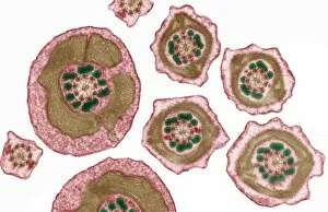
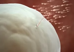
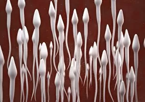
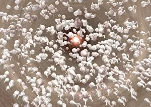
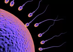
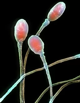
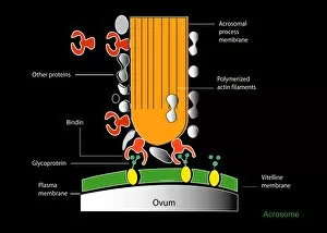
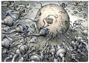
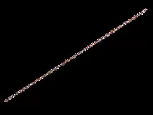
"Spermatozoa: The Tiny Warriors of Life" In the vast world of reproductive biology, spermatozoa play a pivotal role in the creation of life. These microscopic cells hold immense power and are often depicted as warriors on a mission to fertilize an egg. Picture No. 11675522 showcases the intricate dance between an IUD contraceptive and sperm cells, representing the constant battle for conception that occurs within our bodies. This image serves as a reminder of how these resilient little beings persistently strive towards their ultimate goal. Frozen sperm, conceptual image (Picture No. 11675577) highlights the advancements in modern science that allow us to preserve these vital cells for future use. It symbolizes hope for couples struggling with infertility or those who wish to plan their families at a later stage. Microscopic views of human spermatozoa in semen (Picture No. 11675576) provide us with an up-close look at these remarkable creatures swimming through their natural habitat – semen. Their unique shape and tail propulsion system enable them to navigate this complex environment with precision and determination. Human sperm cells (Picture No. 11675524) capture the essence of life's beginnings - each cell carrying half of our genetic material, ready to unite with an egg and create new life. Illustration showing the life cycle of a fern, inset displaying spermatozoa entering female organ (archegonia), emphasizes that even beyond humans, these tiny warriors exist across various species, playing crucial roles in reproduction (Picture No. 11675523). The remaining pictures - Picture Nos. 11675521-20 - further emphasize different aspects related to spermatozoa; they represent diverse perspectives on this fascinating subject matter.