
Spore Bearing Collection
"Exploring the Fascinating World of Spore Bearing: RBG18-3491" Delve into the intriguing realm with RBG18-3491, where ancient fossils and microscopic wonders await


For sale as Licensed Images
Choose your image, Select your licence and Download the media
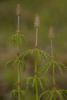
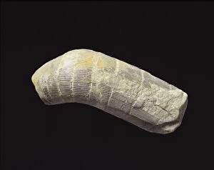
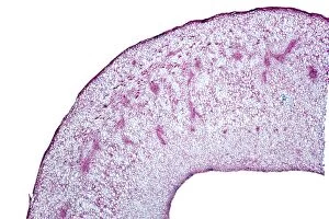
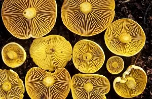
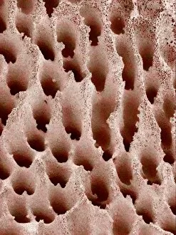
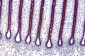
"Exploring the Fascinating World of Spore Bearing: RBG18-3491" Delve into the intriguing realm with RBG18-3491, where ancient fossils and microscopic wonders await. Witness the secrets hidden within Cooksonia plant fossil C013 / 6542 as it reveals its spore-bearing structures from a bygone era. Marvel at the preserved beauty of Fossil horsetail (Calamites sp. ) trunk C013 / 6541, showcasing its intricate spores that once thrived millions of years ago. Zooming in closer, discover the captivating Plum pocket infection through a light micrograph, unveiling an enchanting world teeming with tiny spores. Take a journey to explore Mushroom surface through scanning electron microscopy (SEM), revealing its textured landscape adorned with countless life-giving spores. Venturing further into nature's diversity, encounter vibrant Sulphur toadstools standing tall amidst their surroundings, spreading their reproductive magic through delicate yet resilient spores. Observe Bracken spores under SEM - these minuscule marvels hold immense potential for new life and growth. Uncover the secret language of Fern spores as they communicate their existence under high-resolution SEM imaging. Dive deeper into Mushroom surface through SEM once again; this time exploring every nook and cranny that contributes to its awe-inspiring reproductive capabilities. Switching gears, witness Bracken leaves like never before – magnified by SEM – displaying intricate patterns that aid in efficient dispersal of their precious cargo: thousands upon thousands of tiny yet mighty bracken fern spores. Finally, get up close and personal with Lichen via SEM imagery; this symbiotic masterpiece showcases how two organisms work together harmoniously while producing abundant quantities of resilient fungal or algal-spored offspring. RBG18-3491 invites you on an extraordinary journey filled with ancient fossils and microscopic wonders - a captivating exploration of the diverse and intricate world of spore bearing.