
Stenosis Collection
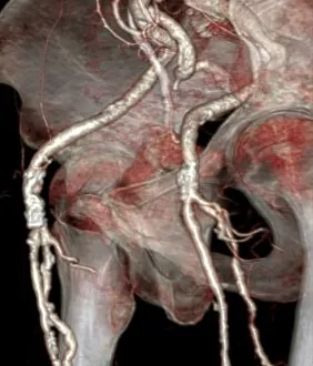
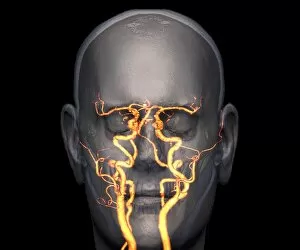
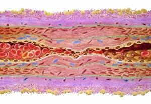
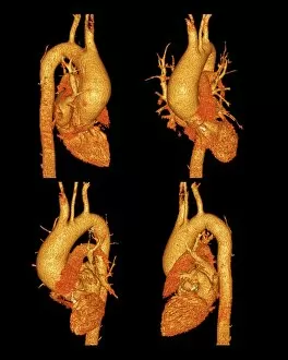
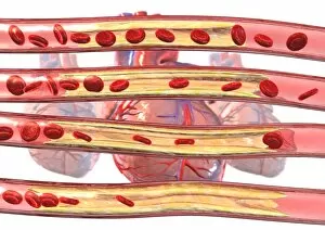
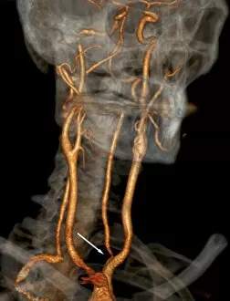
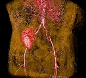
Stenosis, a condition characterized by the narrowing of blood vessels or arteries, can have serious implications for one's health


For sale as Licensed Images
Choose your image, Select your licence and Download the media
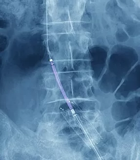
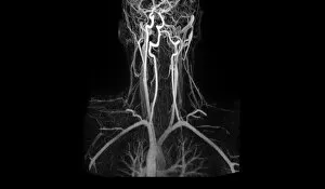
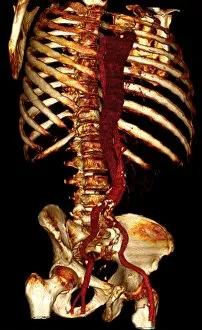
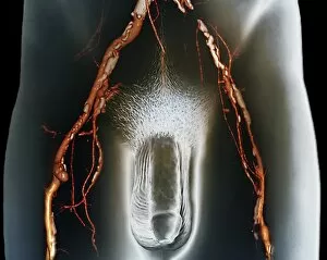
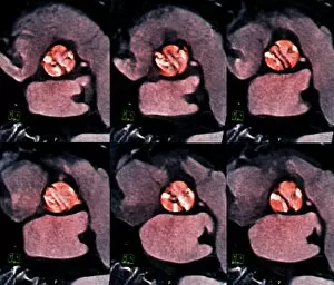
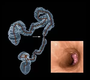
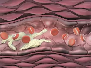
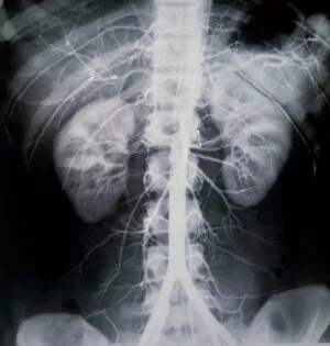
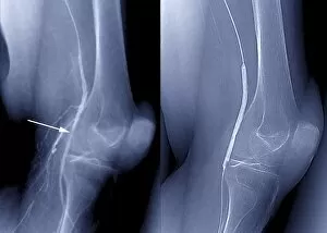
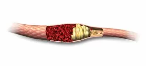
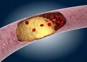
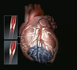
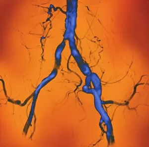
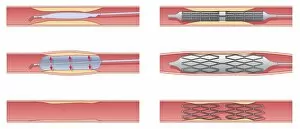
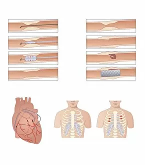
Stenosis, a condition characterized by the narrowing of blood vessels or arteries, can have serious implications for one's health. However, advancements in medical technology and treatments offer hope for those affected. Balloon angioplasty is a common procedure used to treat stenosis. By inserting a deflated balloon into the blocked artery and inflating it, doctors can widen the vessel and restore proper blood flow. X-ray images help guide them during this intricate process. Artery damage caused by they are be detected through an MRA scan. This non-invasive imaging technique provides detailed pictures of blood vessels, allowing doctors to identify any abnormalities or blockages that may require intervention. Computer artwork helps visualize the impact on our blood vessel anatomy. It demonstrates how narrowed arteries due to plaque buildup restrict the smooth flow of oxygen-rich blood throughout our bodies. In cases where stenosis affects larger arteries like the aorta, 3D CT scans are utilized to diagnose conditions such as aortic dissection. These scans provide three-dimensional images that aid in accurate diagnosis and treatment planning. Before treatment for coronary stenosis begins, X-rays play a crucial role in assessing its severity. They reveal areas of blockage within coronary arteries before interventions like angioplasty or bypass surgery are performed. Post-treatment X-rays show remarkable improvements after addressing coronary stenosis with procedures like balloon angioplasty or placement of stents. The once-narrowed arteries now appear widened and restored, ensuring better circulation and reducing risks associated with heart disease. Understanding these diagnostic tools and treatment options empowers both patients and healthcare professionals alike in their fight against stenosis—a condition that threatens cardiovascular health but can be effectively managed through timely interventions.