
Surgical Collection (#7)

"Surgical Marvels: From UFOs to Cattle Mutilation

Background imageSurgical Collection: Decorative lancet case, circa 1800 C017 / 3572

Decorative lancet case, circa 1800 C017 / 3572
Silver lancet case with a floral decoration. Lancets are double-edged surgical blades with a pointed end. These instruments were used historically in medicine to open patients veins for bloodletting

Background imageSurgical Collection: Lancets with cases, circa 1790 -1830 C017 / 3578

Lancets with cases, circa 1790 -1830 C017 / 3578
Two lancets with their cases. The smaller lancet (left) dates from around 1790 and the larger (right), from around 1830. Both lancets are English and made from steel (blade) and tortoiseshell (handle)

Background imageSurgical Collection: Back pain, artwork F007 / 8390

Back pain, artwork F007 / 8390
Back pain, computer artwork

Background imageSurgical Collection: Back pain, artwork F007 / 8389

Back pain, artwork F007 / 8389
Back pain, computer artwork

Background imageSurgical Collection: Back pain, artwork F007 / 8387

Back pain, artwork F007 / 8387
Back pain, computer artwork

Background imageSurgical Collection: Back pain, artwork F007 / 8388

Back pain, artwork F007 / 8388
Back pain, computer artwork

Background imageSurgical Collection: Medical equipment and drugs F008 / 3189

Medical equipment and drugs F008 / 3189
Medical equipment and drugs

Background imageSurgical Collection: Medical equipment and drugs F008 / 3190

Medical equipment and drugs F008 / 3190
Medical equipment and drugs

Background imageSurgical Collection: Medical equipment and drugs F008 / 3188

Medical equipment and drugs F008 / 3188
Medical equipment and drugs

Background imageSurgical Collection: Oro-antral communication, artwork

Oro-antral communication, artwork
Oro-antral communication. Artwork of a human head showing a missing tooth that has developed into a passageway ( communication ) between the mouth (oral cavity) and the maxillary sinus (antrum, red)

Background imageSurgical Collection: Re-enactment of first anaesthesia, 1850

Re-enactment of first anaesthesia, 1850
Re-enactment of first anaesthesia. Group of surgeons and a patient re-enacting the first operation that was carried out under general anaesthesia

Background imageSurgical Collection: Joseph Lister, British surgeon

Joseph Lister, British surgeon
Joseph Lister (1827-1912), British surgeon. Lister qualified as a surgeon in London in 1852. At that time surgery was usually followed by infection and high mortality rates

Background imageSurgical Collection: Cavus foot surgery, ankle tendons

Cavus foot surgery, ankle tendons
Cavus foot surgery. Close-up of surgeons cutting the ankles peroneus tendon before attaching it to another tendon. This is being done during cavus foot surgery

Background imageSurgical Collection: Cavus foot surgery, metatarsal pinning

Cavus foot surgery, metatarsal pinning
Cavus foot surgery. Close-up of bone drilling equipment being used to drill a hole in a metatarsal bone in a patients foot in a case of cavus foot surgery

Background imageSurgical Collection: Toe joint replacement surgery

Toe joint replacement surgery. Close-up of surgeons using a bone saw during an operation to replace a joint in the second toe of a patients foot. For the joint replacement, see image C014/7787

Background imageSurgical Collection: Lancet case with lancets, circa 1800 C017 / 3587

Lancet case with lancets, circa 1800 C017 / 3587
Silver lancet case containing tortoiseshell and steel lancets. Lancets are double edged surgical blades with a pointed end. These instruments were used to open patients veins for bloodletting

Background imageSurgical Collection: Two lancets and their case, circa 1790 C017 / 3560

Two lancets and their case, circa 1790 C017 / 3560
Two lancets with tortoiseshell handles and their silver and fishskin case. These were made in England by Simpson of the Strand around the year 1790

Background imageSurgical Collection: Mechanical scarificators, 19th century C017 / 3564

Mechanical scarificators, 19th century C017 / 3564
Two cased single-bladed mechanical scarificators made from brass and steel. Instruments such as these were designed to create wounds on the surface of the skin

Background imageSurgical Collection: Cupping set, 19th century C017 / 3590

Cupping set, 19th century C017 / 3590
Cupping set dating from around the year 1860, England. This set comprises of four cups and two brass and steel scarificators, complete with a morocco leather case

Background imageSurgical Collection: Cupping set, 19th century C017 / 3569

Cupping set, 19th century C017 / 3569
Cupping set dating from 1880, France. This set comprises of four cups, a syringe, and a scarificator. This set would have been used in a procedure called wet cupping

Background imageSurgical Collection: Mechanical scarificators, circa 1780-1900 C017 / 3562

Mechanical scarificators, circa 1780-1900 C017 / 3562
A collection of single and multibladed mechanical scarificators. These include examples from England, France and Germany and they date from between 1780 and 1900

Background imageSurgical Collection: Ankle surgery: dissecting out tissues C014 / 7781

Ankle surgery: dissecting out tissues C014 / 7781
Ankle surgery. Close-up of surgical tools being used to dissect tissue from a patients ankle. The tool at right is a bone saw, with retractors being used to keep the surgical incision open

Background imageSurgical Collection: Silver medical instruments, 19th century C017 / 0742

Silver medical instruments, 19th century C017 / 0742
Collection of miscellaneous medical silver, including tongue scrapers, sick syphon, nipple shield and lancet case. These instruments are English and date from between 1800 and 1860

Background imageSurgical Collection: Six blade scarificator, 20th century C017 / 3571

Six blade scarificator, 20th century C017 / 3571
Six blade scarificator, made in France around the year 1900. This instrument is made from steel and was designed to create wounds on the surface of the skin, principally for wet cupping procedures

Background imageSurgical Collection: Surgical Instruments. C017 / 3470

Surgical Instruments. C017 / 3470
Plate from 18th century encyclopedia showing illustrations of various surgical instruments. A surgical instrument is a specially designed tool or device for performing specific actions such as

Background imageSurgical Collection: Silver etui with 3 lancets, 18th century C017 / 3591

Silver etui with 3 lancets, 18th century C017 / 3591
Silver etui engraved with a lion design and three lancets. Lancets are double edged surgical blades with a pointed end. These instruments were used to open patients veins for bloodletting

Background imageSurgical Collection: Scarificators and blades, circa 1800 C017 / 3559

Scarificators and blades, circa 1800 C017 / 3559
Three multi-bladed scarificators, with spare blades. Scarificators such as these were used to create wounds on the surface of the skin, principally for wet cupping procedures

Background imageSurgical Collection: Mr. John Taylors cupping set, circa 1870

Mr. John Taylors cupping set, circa 1870
Cupping set, formerly owned by Mr. John Taylor, obstetrician to Winston Churchills mother. In wet cupping procedures, a vacuum created within glass cups would be used to suck blood out of wounds made

Background imageSurgical Collection: Ventricular shunt, X-ray

Ventricular shunt, X-ray
Ventricular shunt. X-ray of the head of a 52-year-old patient with hydrocephalus, showing the position of a ventricular shunt (dark lines, right) and its external valve (round, centre right)

Background imageSurgical Collection: Total knee replacement, X-ray

Total knee replacement, X-ray
Total knee replacement. X-ray of the legs of a 49-year-old patient who has had a total knee replacement (white) of the left knee (right)

Background imageSurgical Collection: Prosthetic hip joint and Gruen zones C016 / 6778

Prosthetic hip joint and Gruen zones C016 / 6778
Prosthetic hip joint and Gruen zones. Cutaway diagram of a femur (thigh bone) showing a femoral component of a hip prosthesis and labels indicating Gruen zones

Background imageSurgical Collection: Prosthetic hip joints and Gruen zones C016 / 6781

Prosthetic hip joints and Gruen zones C016 / 6781
Prosthetic hip joints and Gruen zones. Diagram of the femoral components of two types of hip prosthesis with labels indicating standard and modified Gruen zones

Background imageSurgical Collection: Lumbar spine fusion, artwork C016 / 6993

Lumbar spine fusion, artwork C016 / 6993
Lumbar spine fusion. Artwork of pins and screws inserted into a patients lumbar spine in a case of spinal fusion. The pins hold together the patients vertebrae

Background imageSurgical Collection: Prosthetic hip joint, artwork C016 / 6782

Prosthetic hip joint, artwork C016 / 6782
Prosthetic hip joint. Cutaway artwork of a femur (thigh bone) showing a femoral component of a hip prosthesis. This component is implanted in the femur after the head of the femur has been surgically

Background imageSurgical Collection: Bone drill and grafter, artwork C016 / 6845

Bone drill and grafter, artwork C016 / 6845
Bone drill and grafter. Artwork of a cross-section through a bone drill and grafter that is being used in a hip socket bone grafting operation

Background imageSurgical Collection: Biliary drainage, X-ray C016 / 6558

Biliary drainage, X-ray C016 / 6558
Biliary drainage. Coloured X-ray of the abdomen of an 69 year old patient undergoing biliary drainage. This is a procedure to drain a blocked bile duct

Background imageSurgical Collection: Hip socket bone grafting, diagram C016 / 6786

Hip socket bone grafting, diagram C016 / 6786
Hip socket bone grafting. Diagram showing completed hip socket bone grafting as part of a total hip replacement operation

Background imageSurgical Collection: Hip socket bone grafting, diagram C016 / 6784

Hip socket bone grafting, diagram C016 / 6784
Hip socket bone grafting. Diagram showing the second stage in hip socket bone grafting as part of a total hip replacement operation

Background imageSurgical Collection: Knee osteotomy, artwork C016 / 6995

Knee osteotomy, artwork C016 / 6995
Knee osteotomy. Artwork of a bone tool being used during an osteotomy operation on the knee. An osteotomy involves the removal of bone

Background imageSurgical Collection: Neck arthritis treatment, angiogram C016 / 6440

Neck arthritis treatment, angiogram C016 / 6440
Neck arthritis treatment. Coloured X-ray angiogram of the neck of a 48 year old patient with severe osteoarthritis of the neck

Background imageSurgical Collection: Mitral valve annuloplasty, artwork C016 / 6526

Mitral valve annuloplasty, artwork C016 / 6526
Mitral valve annuloplasty. Artwork of the hearts mitral valve and the coronary sinus (purple), with a stent (metal rod and mesh) inserted into the latter to correct leaking of the mitral valve

Background imageSurgical Collection: Neck arthritis treatment, angiogram C016 / 6443

Neck arthritis treatment, angiogram C016 / 6443
Neck arthritis treatment. Coloured X-ray angiogram of the neck of a 48 year old patient with severe osteoarthritis of the neck

Background imageSurgical Collection: Neck arthritis treatment, X-ray C016 / 6439

Neck arthritis treatment, X-ray C016 / 6439
Neck arthritis treatment. Coloured X-ray of the neck of a 48 year old patient with severe osteoarthritis of the neck, whose cervical spine has been stabilised with a metal plate (rectangular)

Background imageSurgical Collection: Hip socket bone grafting, diagram C016 / 6785

Hip socket bone grafting, diagram C016 / 6785
Hip socket bone grafting. Diagram showing the third stage in hip socket bone grafting as part of a total hip replacement operation

Background imageSurgical Collection: Hip socket bone grafting, diagram C016 / 6783

Hip socket bone grafting, diagram C016 / 6783
Hip socket bone grafting. Diagram showing the first stage in hip socket bone grafting as part of a total hip replacement operation

Background imageSurgical Collection: Biliary drainage, X-ray C016 / 6547

Biliary drainage, X-ray C016 / 6547
Biliary drainage. Coloured X-ray of the abdomen of an 82 year old patient undergoing biliary drainage. This is a procedure to drain a blocked bile duct

Background imageSurgical Collection: Mitral valve annuloplasty, artwork C016 / 6527

Mitral valve annuloplasty, artwork C016 / 6527
Mitral valve annuloplasty. Artwork of the hearts mitral valve and the coronary sinus (purple), with a stent (metal rod and mesh) inserted into the latter to correct leaking of the mitral valve

Background imageSurgical Collection: Vacuum treatment for ulcer, artwork C016 / 6570

Vacuum treatment for ulcer, artwork C016 / 6570
Vacuum treatment for ulcer. Artwork of an advanced vacuum-assisted technique to treat an ulcer during open-chest surgery. An ulcer is a localised area of tissue degeneration


For sale as Licensed Images

Choose your image, Select your licence and Download the media

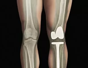
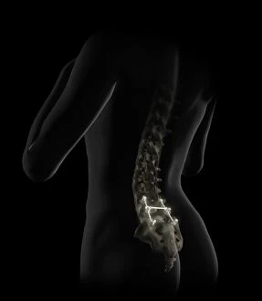
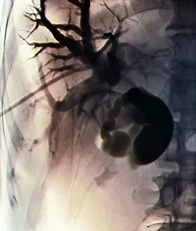
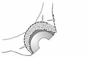
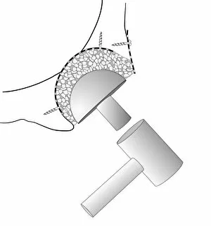
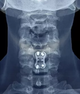
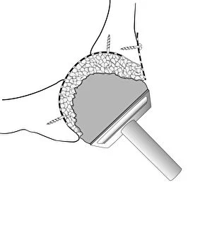
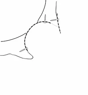
"Surgical Marvels: From UFOs to Cattle Mutilation, Unveiling the Intriguing World of Medical Advancements" Step into the fascinating world wonders as we explore a myriad of captivating hints that have shaped the field throughout history. Delve into the enigmatic realm where Ufos and cattle mutilation intertwine with Joseph Gray and Sons, Surgical Instrument Makers, Truss Works and Star Works on Boston Street - an intriguing trade card from 1890 that leaves us pondering their connection. Travel back in time to the 14th century through trepanation's hauntingly beautiful artwork, showcasing ancient techniques used for cranial surgery. Witness nurses closely observing surgeries, their unwavering dedication evident in every watchful gaze. Marvel at X-ray images revealing total hip replacements, a testament to modern medicine's ability to restore mobility and improve lives. Journey further into medical history with Calots spinal surgery in the 19th century; a groundbreaking procedure that revolutionized treatment for spinal conditions. Behold Goetz von Berlichingen's artificial hand – an extraordinary creation embodying both artistry and functionality. Satirical artwork depicting barber-surgeons reminds us of how far we've come from those unconventional practices. Discover Hugh Owen Thomas, a Welsh surgeon whose contributions continue to shape orthopedic care today. His legacy lives on through his revolutionary methods that transformed patient outcomes forever. Witness heartwarming moments between sister and patient at Convalescent Police Seaside Home in Hove – a reminder of compassion amidst healing journeys. Conceptual artwork unveils eye surgeries' intricate nature while Pasteurs Jubilee celebrations transport us to an era celebrating scientific breakthroughs. Join us as we unravel these surgical mysteries – each hint offering glimpses into humanity's relentless pursuit of knowledge and innovation within this awe-inspiring field.