
Telophase Collection
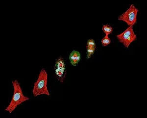
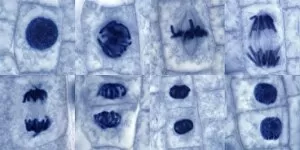
Telophase, the final stage of mitosis, is a captivating process that can be observed through light micrographs and scanning electron micrographs


For sale as Licensed Images
Choose your image, Select your licence and Download the media
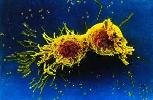
Telophase, the final stage of mitosis, is a captivating process that can be observed through light micrographs and scanning electron micrographs. In plant cell mitosis, a mesmerizing light micrograph showcases the intricate details as the cell divides into two daughter cells. Similarly, in dividing cancer cells captured by SEM C014 / 0362, we witness the intense activity during telophase as these abnormal cells undergo division. The significance becomes even more apparent when examining mouth cancer cells dividing under SEM. The repeated images of mouth cancer cell division emphasize the urgency to understand this process for potential treatments and prevention strategies. Through SEM C014 / 0361, another striking image reveals a dividing cancer cell in its most vulnerable state during telophase. This microscopic view provides valuable insights into cellular mechanisms involved in tumor growth and progression. Mitosis itself is an awe-inspiring phenomenon where one parent cell gives rise to two genetically identical daughter cells. It encompasses several stages including prophase, metaphase, anaphase, and finally culminates in telophase - a crucial step ensuring accurate distribution of genetic material. Scanning electron micrographs allow us to delve deeper into the intricacies of cell division during telophase. These high-resolution images provide scientists with invaluable information about cellular structures and processes at such a minuscule scale.