
Thrombocyte Collection

Thrombocytes, also known as platelets, play a crucial role in our blood clotting process

Background imageThrombocyte Collection: Blood clot, SEM

Blood clot, SEM
Blood clot, coloured scanning electron micrograph (SEM). Red blood cells are red and fibrin protein strands are green. Platelets are at bottom right

Background imageThrombocyte Collection: Blood clot, SEM C016 / 9747

Blood clot, SEM C016 / 9747
Blood clot, coloured scanning electron micrograph (SEM). Red blood cells (erythrocytes) are trapped within a fibrin protein mesh (beige)

Background imageThrombocyte Collection: Lung tissue, TEM

Lung tissue, TEM
Lung tissue, coloured transmission electron micrograph (TEM). A single alveolus is at center. At top right is a pulmonary capillary containing a red blood cell (red) and a platelet (yellow)

Background imageThrombocyte Collection: Conceptual image of platelet, red blood cell and white blood cell

Conceptual image of platelet, red blood cell and white blood cell

Background imageThrombocyte Collection: Blood clot, artwork C016 / 4619

Blood clot, artwork C016 / 4619
Blood clot in a blood vessel, computer artwork. Red blood cells (erythrocytes) are trapped within a fibrin protein mesh (cream)

Background imageThrombocyte Collection: White blood cells and platelets, SEM C016 / 3099

White blood cells and platelets, SEM C016 / 3099
White blood cells and platelets. Coloured scanning electron micrograph (SEM) of platelets (green) with a variety of different white blood cells (leucocytes, yellow)

Background imageThrombocyte Collection: White blood cells and platelets, SEM C016 / 3098

White blood cells and platelets, SEM C016 / 3098
White blood cells and platelets. Coloured scanning electron micrograph (SEM) of platelets (purple) with a variety of different white blood cells (leucocytes, blue)

Background imageThrombocyte Collection: Blood platelets

Blood platelets. Coloured scanning electron micro- graph (SEM) of activated blood platelets. Platelet cells (thrombocytes) are formed in the bone marrow

Background imageThrombocyte Collection: Conceptual image of a group of platelets

Conceptual image of a group of platelets

Background imageThrombocyte Collection: Blood vessel with platelets, white blood cells and red blood cells

Blood vessel with platelets, white blood cells and red blood cells

Background imageThrombocyte Collection: Normal artery compared to plaque and thrombus formation in artery

Normal artery compared to plaque and thrombus formation in artery

Background imageThrombocyte Collection: Conceptual image of platelets with red blood cells

Conceptual image of platelets with red blood cells
Conceptual image of platelets with white blood cells and red blood cells

Background imageThrombocyte Collection: Conceptual image of malaria parasites within red blood cells

Conceptual image of malaria parasites within red blood cells

Background imageThrombocyte Collection: Conceptual image of a platelet

Conceptual image of a platelet

Background imageThrombocyte Collection: Artery showing atherosclerotic plaque, platelets and red blood cells

Artery showing atherosclerotic plaque, platelets and red blood cells
Close-up of artery showing atherosclerotic plaque, platelets and red blood cells

Background imageThrombocyte Collection: Conceptual image of red blood cells with platelets

Conceptual image of red blood cells with platelets

Background imageThrombocyte Collection: Blood clot, SEM C016 / 9751

Blood clot, SEM C016 / 9751
Blood clot, coloured scanning electron micrograph (SEM). Red blood cells (erythrocytes) are trapped within a fibrin protein mesh (beige)

Background imageThrombocyte Collection: Blood clot, SEM C016 / 9746

Blood clot, SEM C016 / 9746
Blood clot, coloured scanning electron micrograph (SEM). Red blood cells (erythrocytes) are trapped within a fibrin protein mesh (beige)

Background imageThrombocyte Collection: Blood clot, SEM C016 / 9749

Blood clot, SEM C016 / 9749
Blood clot, coloured scanning electron micrograph (SEM). Red blood cells (erythrocytes) are trapped within a fibrin protein mesh (beige)

Background imageThrombocyte Collection: Blood clot, SEM C017 / 7141

Blood clot, SEM C017 / 7141
Blood clot, coloured scanning electron micrograph (SEM). Red blood cells (erythrocytes) and a white blood cell (purple) trapped within a fibrin protein mesh (cream)

Background imageThrombocyte Collection: Blood clot, SEM C016 / 9750

Blood clot, SEM C016 / 9750
Blood clot, coloured scanning electron micrograph (SEM). Red blood cells (erythrocytes) are trapped within a fibrin protein mesh (beige)

Background imageThrombocyte Collection: Blood clot, SEM C016 / 9753

Blood clot, SEM C016 / 9753
Blood clot. Coloured scanning electron micrograph (SEM) of a blood clot from the inner wall of the left ventricle of a heart

Background imageThrombocyte Collection: Blood clot, SEM C016 / 9752

Blood clot, SEM C016 / 9752
Blood clot, coloured scanning electron micrograph (SEM). Red blood cells (erythrocytes) are trapped within a fibrin protein mesh (beige)

Background imageThrombocyte Collection: Platelets, artwork F007 / 7458

Platelets, artwork F007 / 7458
Platelets, computer artwork

Background imageThrombocyte Collection: Platelets, artwork F007 / 7459

Platelets, artwork F007 / 7459
Platelets, computer artwork

Background imageThrombocyte Collection: Blood clot, SEM C016 / 9745

Blood clot, SEM C016 / 9745
Blood clot, coloured scanning electron micrograph (SEM). Red blood cells (erythrocytes) are trapped within a fibrin protein mesh (beige)

Background imageThrombocyte Collection: White blood cell and platelet, SEM F006 / 9929

White blood cell and platelet, SEM F006 / 9929
White blood cells. Coloured scanning electron micrograph (SEM) of white blood cells (leucocytes) and a platelet (thrombocyte, red). Magnification: x6, 600 when printed at 10 centimetres wide

Background imageThrombocyte Collection: White blood cell and platelet, SEM F006 / 9928

White blood cell and platelet, SEM F006 / 9928
White blood cells. Coloured scanning electron micrograph (SEM) of white blood cells (leucocytes) and a platelet (thrombocyte, orange). Magnification: x6, 600 when printed at 10 centimetres wide

Background imageThrombocyte Collection: White blood cell and platelet, SEM F006 / 9927

White blood cell and platelet, SEM F006 / 9927
White blood cells. Coloured scanning electron micrograph (SEM) of white blood cells (leucocytes) and a platelet (thrombocyte, green). Magnification: x6, 600 when printed at 10 centimetres wide

Background imageThrombocyte Collection: Blood clot, SEM C016 / 9748

Blood clot, SEM C016 / 9748
Blood clot, coloured scanning electron micrograph (SEM). Red blood cells (erythrocytes) are trapped within a fibrin protein mesh (beige)

Background imageThrombocyte Collection: Platelet, SEM C016 / 9060

Platelet, SEM C016 / 9060
Platelet, coloured scanning electron micrograph (SEM). Platelets, or thrombocytes, are fragments of white blood cells. When blood clots

Background imageThrombocyte Collection: Platelet, SEM C016 / 9059

Platelet, SEM C016 / 9059
Platelet, coloured scanning electron micrograph (SEM). Platelets, or thrombocytes, are fragments of white blood cells. When blood clots

Background imageThrombocyte Collection: Blood clot, SEM C015 / 9608

Blood clot, SEM C015 / 9608
Blood clot. Coloured scanning electron micrograph (SEM) of a blood clot from the inner wall of the left ventricle of a heart

Background imageThrombocyte Collection: Blood cells, illustration C018 / 0802

Blood cells, illustration C018 / 0802
Blood cells. All cellular blood components originate from the same cell, the haematopoietic stem cell. The stem cell differentiates into two types of progenitor cells

Background imageThrombocyte Collection: Blood clot, SEM C016 / 9744

Blood clot, SEM C016 / 9744
Blood clot, coloured scanning electron micrograph (SEM). Red blood cells (erythrocytes) are trapped within a fibrin protein mesh (beige)

Background imageThrombocyte Collection: Blood clot, SEM C014 / 0381

Blood clot, SEM C014 / 0381
Blood clot. Coloured scanning electron micrograph (SEM) of a blood clot (thrombus) in an arteriole (small blood vessel) of a salivary gland

Background imageThrombocyte Collection: Blood clot, SEM C014 / 0380

Blood clot, SEM C014 / 0380
Blood clot. Coloured scanning electron micrograph (SEM) of a blood clot (thrombus) in an arteriole (small blood vessel) of a salivary gland

Background imageThrombocyte Collection: Blood clot, artwork C016 / 4618

Blood clot, artwork C016 / 4618
Blood clot, computer artwork. Red blood cells (erythrocytes) are trapped within a fibrin protein mesh (cream). The fibrin mesh is formed in response to chemicals secreted by platelets (white)

Background imageThrombocyte Collection: Blood clot, artwork C016 / 4620

Blood clot, artwork C016 / 4620
Fortification agate. Section through a fortification agate from Scurdie Ness, Scotland, UK. Agate is an aggregate of different forms of silica (SiO2) within a geode

Background imageThrombocyte Collection: White blood cells, SEM C016 / 3102

White blood cells, SEM C016 / 3102
White blood cells. Coloured scanning electron micrograph (SEM) of white blood cells (leucocytes) from a sample of peripheral blood

Background imageThrombocyte Collection: Blood cells, SEM C016 / 3101

Blood cells, SEM C016 / 3101
Blood cells. Coloured scanning electron micrograph (SEM) of a variety of different white blood cells (leucocytes, blue/purple) with two red blood cells (erythrocytes)

Background imageThrombocyte Collection: Blood cells, SEM C016 / 3100

Blood cells, SEM C016 / 3100
Blood cells. Coloured scanning electron micrograph (SEM) of a variety of different white blood cells (leucocytes, yellow/green) with two red blood cells (erythrocytes)

Background imageThrombocyte Collection: Macrophage and platelets, SEM C016 / 3094

Macrophage and platelets, SEM C016 / 3094
Macrophage and platelets. Coloured scanning electron micrograph (SEM) of platelets (green) attached to a macrophage white blood cell (turquoise)

Background imageThrombocyte Collection: Platelets, SEM C016 / 3091

Platelets, SEM C016 / 3091
Platelets, coloured scanning electron micrograph (SEM). Platelets, or thrombocytes, are fragments of white blood cells. When blood clots

Background imageThrombocyte Collection: Blood clot C013 / 7129

Blood clot C013 / 7129
Blood clot. Coloured scanning electron micrograph (SEM) of a blood clot (centre) forming in connective tissue. Red blood cells (erythrocytes)

Background imageThrombocyte Collection: Blood clot C013 / 7128

Blood clot C013 / 7128
Blood clot. Coloured scanning electron micrograph (SEM) of a blood clot (centre) forming in connective tissue. Red blood cells (erythrocytes)

Background imageThrombocyte Collection: Blood cells, artwork C013 / 4726

Blood cells, artwork C013 / 4726
Blood cells. Computer artwork of red blood cells (erythrocytes, red), white blood cells (leucocytes, white), and platelets (thrombocytes, yellow)

Background imageThrombocyte Collection: Blood cells, artwork C013 / 4707

Blood cells, artwork C013 / 4707
Blood cells. Computer artwork of red blood cells (erythrocytes, red), white blood cells (leucocytes, white), and platelets (thrombocytes, yellow)


For sale as Licensed Images

Choose your image, Select your licence and Download the media

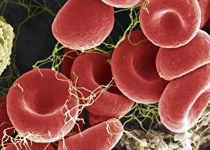
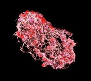
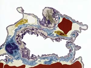
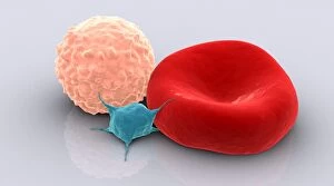
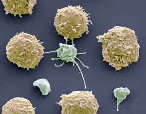
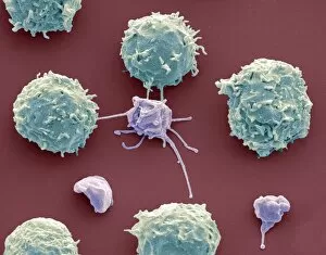
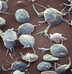
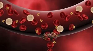
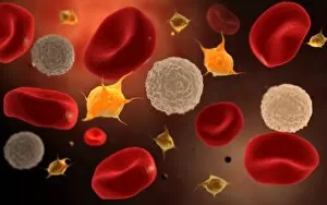
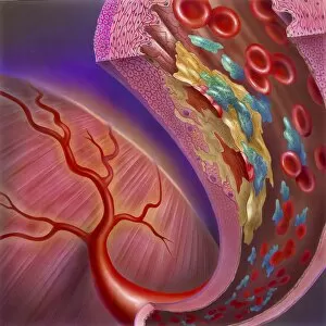
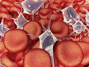
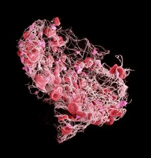
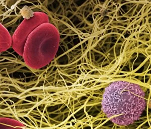
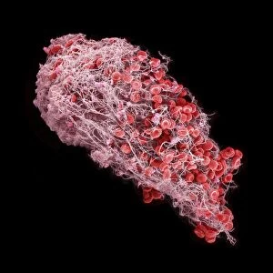
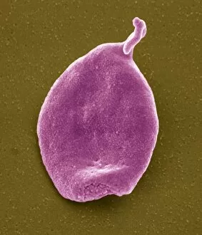
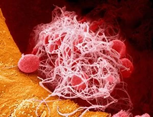
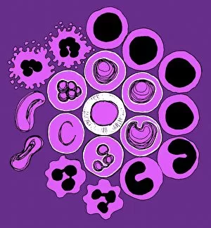
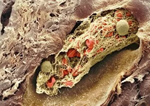
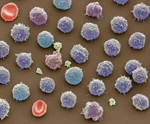
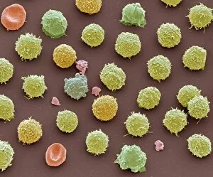
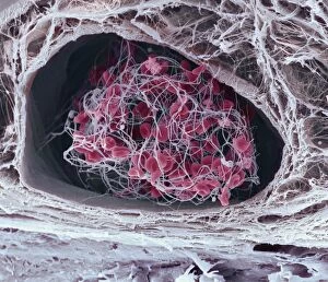
Thrombocytes, also known as platelets, play a crucial role in our blood clotting process. These tiny cell fragments are captured beautifully under the scanning electron microscope (SEM C016 / 9747), showcasing their intricate structure. In this conceptual image, we witness the collaboration between thrombocytes and other blood components like red and white blood cells. It's fascinating to see how these different elements work together to form a protective barrier against bleeding or injury (Platelets, SEM). Another captivating artwork (Blood clot, artwork C016 / 4619) depicts the formation of a blood clot. This natural defense mechanism prevents excessive bleeding by forming a mesh-like structure composed of platelets and fibrin strands. Under SEM observation (White blood cells and platelets, SEM C016 / 3099), we can appreciate the unique characteristics of both white blood cells and platelets. Their distinct shapes and sizes contribute to their specific functions within our immune system. A conceptual image featuring platelets alongside red blood cells highlights their close association during normal physiological processes (Conceptual image of platelets with red blood cells). Moreover, another conceptual image showcases an assembly of these remarkable cell fragments called "a group of platelets, " emphasizing their collective power in maintaining hemostasis. When examining a cross-section of an artery, we can observe the stark contrast between a healthy vessel and one affected by plaque buildup or thrombus formation (Normal artery compared to plaque and thrombus formation in artery). Thrombocytes play an essential role in preventing such complications by swiftly responding to injuries within our circulatory system. These captivating images shed light on the intricate world of thrombocytes – from their microscopic structures seen through powerful microscopes to their vital contributions in safeguarding our health.