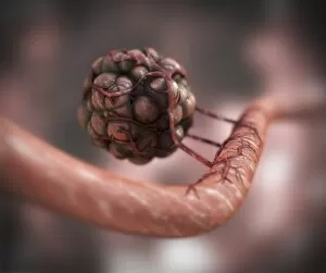
Tumour Collection (page 4)

"Tumour: Unveiling the Battle Within" In a microscopic realm, T lymphocytes engage in an intense combat against cancer cells, as captured by SEM C001 / 1679

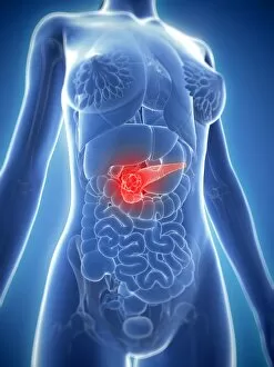
Background imageTumour Collection: Pancreatic cancer, artwork F006 / 7997

Pancreatic cancer, artwork F006 / 7997
Pancreatic cancer, computer artwork

Background imageTumour Collection: Female stomach, artwork

Female stomach, artwork
Female stomach, computer artwork

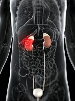
Background imageTumour Collection: Kidney cancer, artwork F006 / 7943

Kidney cancer, artwork F006 / 7943
Kidney cancer, computer artwork

Background imageTumour Collection: Kidney cancer, artwork F006 / 7941

Kidney cancer, artwork F006 / 7941
Kidney cancer, computer artwork

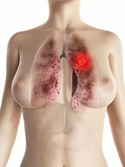
Background imageTumour Collection: Female lung cancer, artwork

Female lung cancer, artwork
Female lung cancer, computer artwork

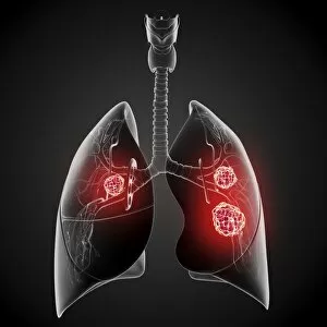
Background imageTumour Collection: Lung cancer, artwork

Lung cancer, artwork
Lung cancer, computer artwork

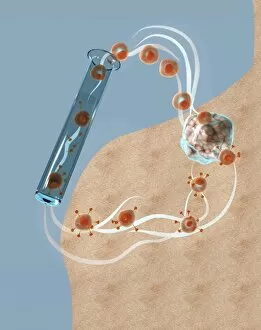
Background imageTumour Collection: Immunotherapy, artwork

Immunotherapy, artwork
Immunotherapy. Computer artwork of immunotherapy being used to destroy a tumour (right). Immunotherapy uses the bodys immune system to fight a disease

Background imageTumour Collection: Cancer cells spreading, artwork

Cancer cells spreading, artwork
Cancer cells spreading. Computer artwork showing cancerous cells (blue) migrating from a tumour (upper centre) into the blood stream (centre)

Background imageTumour Collection: Spinal cancer, MRI scan

Spinal cancer, MRI scan
Spinal cancer. Coloured magnetic resonance imaging (MRI) scan of a sagittal section through the cervical (neck) region of the spine of a 22-year-old patient

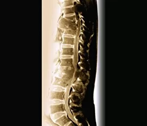
Background imageTumour Collection: Secondary spinal cancer, MRI scan

Secondary spinal cancer, MRI scan
Secondary spinal cancer. Magnetic resonance imaging (MRI) scan of a section through the spine of a 48-year-old patient, showing malignant (cancerous) tumours

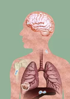
Background imageTumour Collection: Metastatic cancer, artwork

Metastatic cancer, artwork
Metastatic cancer. Computer artwork of a section through a persons upper body and head, showing a primary cancer in the right liver (lower left) that has spread (metastasised)

Background imageTumour Collection: Cancer blood supply, conceptual image C016 / 7505

Cancer blood supply, conceptual image C016 / 7505
Cancer blood supply, conceptual image. Computer artwork representing the blood supply to a cancerous (malignant) tumour

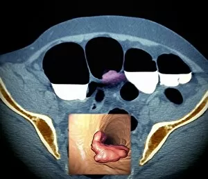
Background imageTumour Collection: Colon cancer, barium contrast CT scan

Colon cancer, barium contrast CT scan
Colon cancer. Coloured barium contrast computed tomography (CT) scan, and 3D CT colonoscopy image (inset), of an axial section through the sigmoid colon (large intestine) of a 54-year-old patient
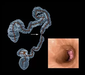
Background imageTumour Collection: Colon cancer, 3D colonoscopy image

Colon cancer, 3D colonoscopy image
Colon cancer. 3D colonoscopy image showing the location of a cancerous growth (centre right) in the colon of a 59-year-old patient. Here, the growth has caused a narrowing (stenosis) of the colon

Background imageTumour Collection: Breast lumps, MRI

Breast lumps, MRI
Breast lumps. Coloured magnetic resonance imaging (MRI) scan of the chest of a 20-year-old patient, showing multiple fibroadenomas (light patches) in both breasts

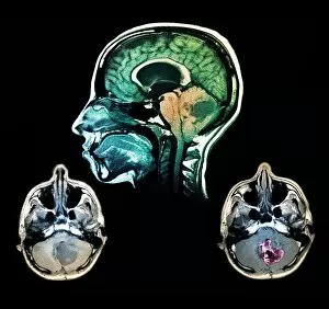
Background imageTumour Collection: Brain tumour, MRI scans

Brain tumour, MRI scans
Brain tumour. Coloured magnetic resonance imaging (MRI) scans of sections through the head of a 23-year-old patient with a lesion (dark patches)

Background imageTumour Collection: Kidney tumour, CT scan

Kidney tumour, CT scan
Kidney tumour. Coloured computed tomography (CT) scan of a section through the abdomen of a 62-year-old patient, showing necrosis (death) of tissue in the right kidney

Background imageTumour Collection: Secondary lung cancer, X-ray

Secondary lung cancer, X-ray
Secondary lung cancers. Coloured X-ray of malignant (cancerous) tumours (red patches) in the lungs of a 60-year-old female patient

Background imageTumour Collection: Colon cancer, CT scans

Colon cancer, CT scans
Colon cancer. 2D computed tomography (CT) rendering (left) and 3D colonoscopy image (right) showing the location of a cancerous growth in the colon of a 59-year-old patient

Background imageTumour Collection: Effect of oxygen supply on tumour growth C016 / 6256

Effect of oxygen supply on tumour growth C016 / 6256
Effect of oxygen supply on tumour growth. Top: a small blood supply (tubes) means a low level of oxygen is available and the tumour (round) grows slowly

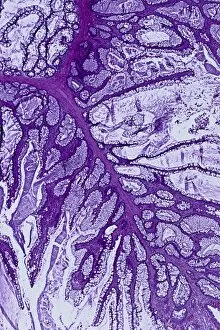
Background imageTumour Collection: Colon polyp, light micrograph C016 / 0512

Colon polyp, light micrograph C016 / 0512
Colon polyp. Light micrograph of a section through part of a tubulovillous adenoma of the mucosa of the large bowel (colon)

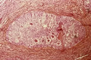
Background imageTumour Collection: Brenner tumour of the ovary C015 / 7137

Brenner tumour of the ovary C015 / 7137
Brenner tumour of the ovary. Light micrograph of a section through an ovary showing a Brenner tumour (centre). This rare benign (non-cancerous)

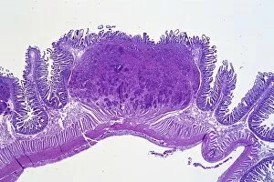
Background imageTumour Collection: Bowel cancer, light micrograph C016 / 0511

Bowel cancer, light micrograph C016 / 0511
Bowel cancer. Light micrograph of a section through a carcinoid tumour in the small bowel. The tumour has invaded the submucosa of the bowel

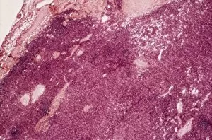
Background imageTumour Collection: Thymus gland cancer, light micrograph C015 / 7128

Thymus gland cancer, light micrograph C015 / 7128
Thymus cancer. Light micrograph of a section through a thymus gland with thymoma cancer. The thymus is a gland in the upper chest that plays an important role in the immune system

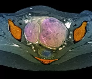
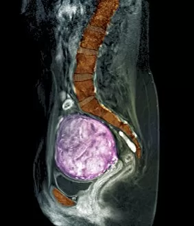
Background imageTumour Collection: Uterine fibroids, MRI scan C018 / 0467

Uterine fibroids, MRI scan C018 / 0467
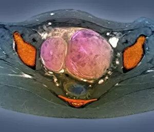
Uterine fibroids. Coloured magnetic resonance imaging (MRI) scan of the pelvis of a 47 year old female with two large fibroids (pink) in her uterus

Background imageTumour Collection: Rectal cancer, MRI scans C018 / 0508

Rectal cancer, MRI scans C018 / 0508
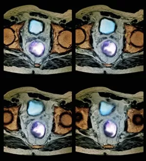
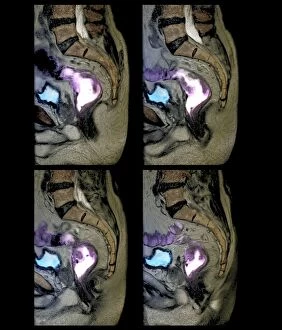
Rectal cancer. Coloured magnetic resonance imaging (MRI) scans of the pelvis of a 65 year old patient with a malignant (cancerous) tumour (purple) in the rectum, the last part of the large intestine

Background imageTumour Collection: Rectal cancer, MRI scans C018 / 0510

Rectal cancer, MRI scans C018 / 0510
Rectal cancer. Coloured magnetic resonance imaging (MRI) scans of the pelvis of a 65 year old patient with a malignant (cancerous) tumour in the rectum, the last part of the large intestine

Background imageTumour Collection: Uterine fibroids, MRI scan C018 / 0468

Uterine fibroids, MRI scan C018 / 0468
Uterine fibroids. Coloured magnetic resonance imaging (MRI) scan of the pelvis of a 47 year old female with two large fibroids (pink) in her uterus

Background imageTumour Collection: Liver cancer, CT scan C018 / 0461

Liver cancer, CT scan C018 / 0461
Liver cancer. Coloured computed tomography (CT) scan of the abdomen of a 50 year old alcoholic patient with liver cancer. The liver (orange) is enlarged due to the presence of the tumour

Background imageTumour Collection: Rectal cancer, MRI scans C018 / 0511

Rectal cancer, MRI scans C018 / 0511
Rectal cancer. Coloured magnetic resonance imaging (MRI) scans of the pelvis of a 65 year old patient with a malignant (cancerous) tumour (purple) in the rectum, the last part of the large intestine

Background imageTumour Collection: Colon cancer, barium X-ray C018 / 0574

Colon cancer, barium X-ray C018 / 0574
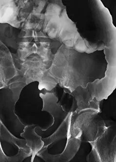
Colon cancer. Coloured X-ray of the large intestine of a 62 year old patient with cancer of the sigmoid colon. The patient was given a barium enema prior to the X-ray to show up any abnormalities

Background imageTumour Collection: Liver cancer, CT scan C018 / 0462

Liver cancer, CT scan C018 / 0462
Liver cancer. Computed tomography (CT) scan of the abdomen of a 50 year old alcoholic patient with liver cancer. The liver (left to upper centre) is enlarged due to the presence of the tumour

Background imageTumour Collection: Uterine fibroids, MRI scan C018 / 0469

Uterine fibroids, MRI scan C018 / 0469
Uterine fibroids. Coloured magnetic resonance imaging (MRI) scan of the pelvis of a 47 year old female with two large fibroids (pink) in her uterus

Background imageTumour Collection: Animal models for cancer research C018 / 0748

Animal models for cancer research C018 / 0748
Animal models for cancer research, conceptual illustration. Fruit fly (Drosophila melanogaster) with malignant (cancerous) tumours in one of its eyes

Background imageTumour Collection: Liver cancer, CT scan C018 / 0460

Liver cancer, CT scan C018 / 0460
Liver cancer. Coloured computed tomography (CT) scan of the abdomen of a 50 year old alcoholic patient with liver cancer. The liver (orange) is enlarged due to the presence of the tumour

Background imageTumour Collection: Rectal cancer, MRI scan C018 / 0512

Rectal cancer, MRI scan C018 / 0512
Rectal cancer. Coloured magnetic resonance imaging (MRI) scan of the pelvis of a 65 year old patient with a malignant (cancerous) tumour in the rectum, the last part of the large intestine

Background imageTumour Collection: Benign dental tumour, CT scans C018 / 0589

Benign dental tumour, CT scans C018 / 0589
Benign dental tumour. Coloured computed tomography (CT) scans of the lower jaw of a 35 year old patient with a cemento-ossifying fibroma (purple)

Background imageTumour Collection: Jaw tumour, CT scan C018 / 0565

Jaw tumour, CT scan C018 / 0565
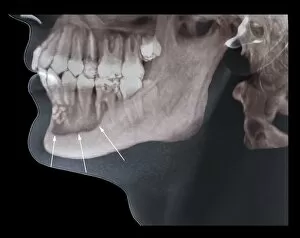
Jaw tumour. Coloured 3D computed tomography (CT) scan of the jaw of a 35 year old patient showing a benign (non-cancerous) bony lesion (indicated by arrows) in the mandible (lower jaw)

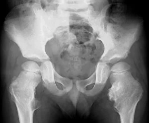
Background imageTumour Collection: Bone growths, X-ray C018 / 0580

Bone growths, X-ray C018 / 0580
Bone growths. X-ray of the pelvis of a 12 year old patient with bony growths (exostoses) and cartilage cysts (enchondromas) on the femurs (thigh bones) and hip bones

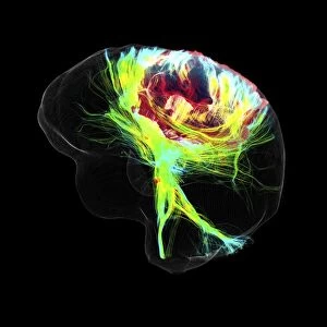
Background imageTumour Collection: Glioblastoma brain tumour, DTI MRI scan C017 / 7052

Glioblastoma brain tumour, DTI MRI scan C017 / 7052
Glioblastoma brain tumour. 3D diffusion tensor imaging (DTI) magnetic resonance imaging (MRI) scan of nerve pathways (coloured) around a glioblastoma (upper right) in the brain

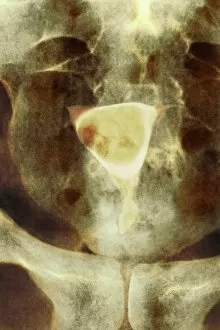
Background imageTumour Collection: Uterine fibroid, X-ray C014 / 4919

Uterine fibroid, X-ray C014 / 4919
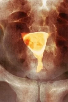
Uterus tumour. Coloured X-ray of the abdomen of a woman with a fibroid (round, centre) in her uterus. A fibroid is a fibrous benign tumour originating from muscular tissue (leimyoma)

Background imageTumour Collection: Uterine fibroid, X-ray C014 / 4918

Uterine fibroid, X-ray C014 / 4918
Uterus tumour. Coloured X-ray of the abdomen of a woman with a fibroid (orange, round) in her uterus. A fibroid is a fibrous benign tumour originating from muscular tissue (leimyoma)

Background imageTumour Collection: Stomach cancer, barium X-ray

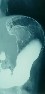
Stomach cancer, barium X-ray
Stomach cancer. Coloured frontal X-ray of the upper gastroduodenal tract of a patient with stomach cancer (grey, upper centre)

Background imageTumour Collection: Intestinal lymphoid hyperplasia C016 / 8334

Intestinal lymphoid hyperplasia C016 / 8334
Intestinal lymphoid hyperplasia. Endoscopic view of lymphoid hyperplasia in the terminal ileum - the last part of the small intestine

Background imageTumour Collection: Hyperplastic polyps in the stomach C016 / 8327

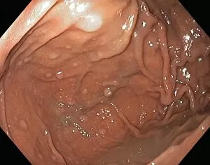
Hyperplastic polyps in the stomach C016 / 8327
Hyperplastic polyps in the stomach. Endoscopic view of small hyperplastic polyps (growths) in the gastric fundus of the stomach

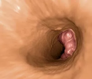
Background imageTumour Collection: Colon cancer, endoscopic view C016 / 8307

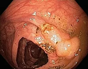
Colon cancer, endoscopic view C016 / 8307
Colon cancer. Endoscopic view of a malignant (cancerous) adenocarcinoma (lower right) in a patients ascending colon (large intestine)

Background imageTumour Collection: Blood vessel formation, artwork

Blood vessel formation, artwork
Blood vessel formation. Artwork showing malignant (cancerous) tumour cells promoting the formation of new blood vessels, a process known as angiogenesis

Background imageTumour Collection: Radiotherapy, conceptual image C013 / 5587

Radiotherapy, conceptual image C013 / 5587
Radiotherapy, conceptual image. Computer artwork representing the use of ionising radiation to kill cancerous cells (centre)


For sale as Licensed Images

Choose your image, Select your licence and Download the media

"Tumour: Unveiling the Battle Within" In a microscopic realm, T lymphocytes engage in an intense combat against cancer cells, as captured by SEM C001 / 1679. These brave warriors of our immune system tirelessly fight to protect us from the clutches of disease. Moving to a different battleground, ovarian cancer reveals its intricate nature under the lens of a light micrograph (C015 / 7103). This visual exploration sheds light on the complexity and urgency surrounding this formidable adversary. Amidst these scientific endeavors, nanorobots emerge as potential allies in our war against cancer. Witness their futuristic assault on malignant cells, offering hope for a brighter future where tumors are eradicated (Nanorobot attacking cancer). Beyond the realm of science lies another facet of tumours - congenital eye anomalies. Klinische Darstellungen der provides insight into these mysterious conditions that affect vision and challenge medical professionals' understanding. Exploring further, we encounter "Kyste sereux cloisonne du cou" and "Lipome de la tete, " captivating colour lithographs that depict unique manifestations of tumours in unexpected places. These artistic representations remind us that even within chaos lies beauty waiting to be discovered. Delving into history's archives brings forth Joanna Southcott, an extraordinary fanatic whose beliefs stirred controversy during her time. Like a metaphorical tumour challenging societal norms, she left an indelible mark on history (Joanna Southcott: An Extraordinary Fanatic, 1822). Artistic expressions also capture the essence of tumours; J-L David's masterpiece Chevignard portrays emotions intertwined with physical affliction caused by these growths. Through art's lens emerges empathy for those affected by such ailments (J-L David / Chevignard). Nature too holds its own share of peculiarities – behold Catasetum callosum with its tumor-lipped or callous appearance.