
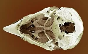
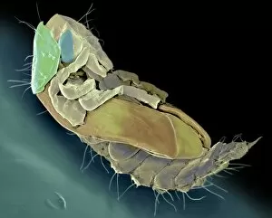
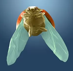
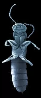
Underneath Collection (#29)
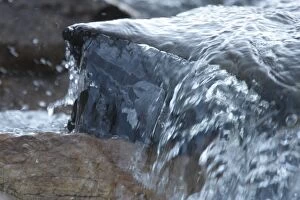
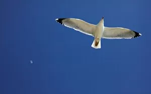
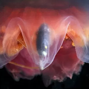
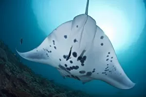
Discovering the Hidden Wonders Underneath: From Cave Hill to Belfast Castle, nature's secrets unfold. Witness the grace of a giant manta ray gliding through the depths


For sale as Licensed Images
Choose your image, Select your licence and Download the media
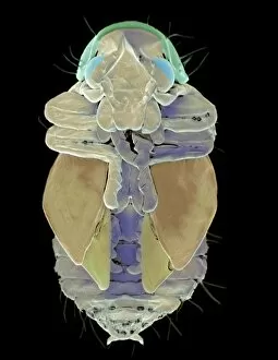
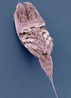
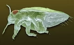
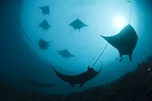
Discovering the Hidden Wonders Underneath: From Cave Hill to Belfast Castle, nature's secrets unfold. Witness the grace of a giant manta ray gliding through the depths. Step back in time to Lambeth Bridge in the 1950s, where nostalgia meets modernity. Explore the Oceanic Pier in Wilmington, North Carolina, and let its charm wash over you. Feel the thrill of holding a stingray in Antigua, amidst the vibrant West Indies' beauty. Dive into an underwater paradise teeming with life at Egypt's Red Sea coral reef – a testament to Earth's immense biodiversity. Marvel at majestic manta rays gracefully dancing beneath turquoise waters. Journey through history as you traverse London's iconic Thames Tunnel or immerse yourself in Lewis Carroll's whimsical world at The Mad Hatters Tea Party scene from c. 1865. Cross Marlow Bridge over Buckinghamshire's serene River Thames and feel tranquility envelop your soul. Admire the power and precision of an F-14A Tomcat armed with missiles soaring high above or witness Ukrainian Air Force Su-27 Flanker showcasing their aerial prowess. From breathtaking natural wonders to captivating man-made marvels, there is always more than meets the eye underneath our world’s surface.