
Upper Jaw Collection
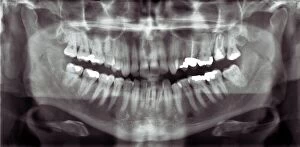
"The Fascinating World of the Upper Jaw: Exploring Dental Anatomy and Procedures" Unveiling the intricate details of dental anatomy with a panoramic dental X-ray

For sale as Licensed Images
Choose your image, Select your licence and Download the media
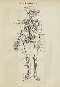
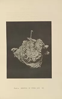
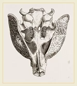
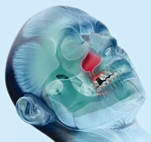
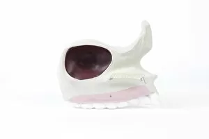
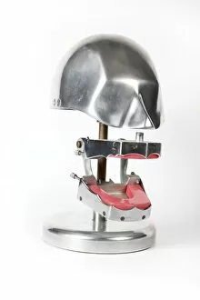
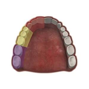
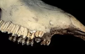
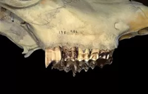
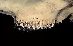
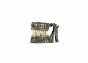
"The Fascinating World of the Upper Jaw: Exploring Dental Anatomy and Procedures" Unveiling the intricate details of dental anatomy with a panoramic dental X-ray. A glimpse into history through an exquisite human skeleton engraving, showcasing the upper jaw. Discovering the pioneering work of Charles B Brigham in removal procedures of the upper jaw in American dentistry. Examining the unique features of a Paca's upper jaw, offering insights into animal dentition. Artistic representation depicting oro-antral communication, highlighting challenges in oral surgery involving the upper jaw. Delving into historical models to understand how our understanding of human upper jaws has evolved over time. Step back in time as we explore vintage dentistry equipment used for various procedures on the upper jaw. Appreciating art's role in educating about nerve blocks with artwork showcasing infraorbital nerve block techniques (C016 / 6818). Understanding anterior superior alveolar nerve block techniques (C016 / 6822) and their significance for pain management during dental procedures on the upper jaw. Exploring posterior superior alveolar nerve block techniques (C016 / 6824) that revolutionized anesthesia during surgeries involving the upper jaw. Diving deeper into dental maxillary nerve regions through captivating artwork (C016 / 6835), unraveling complexities within this vital area related to oral health and sensation. Appreciating art's ability to educate by presenting middle superior alveolar nerve block techniques (C016 / 6823), enhancing our knowledge about anesthesia administration for specific areas within the upper jaw. Intriguingly complex and rich with history, exploring all aspects surrounding our remarkable "upper jaw" reveals its importance not only from a functional perspective but also as an intriguing subject matter intertwining science, medicine, and art throughout centuries past and present.