
Urinary System Collection (#9)
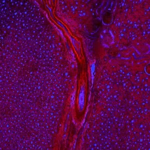
The urinary system is a complex network of organs that play a vital role in maintaining the body's overall health and balance


For sale as Licensed Images
Choose your image, Select your licence and Download the media
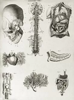
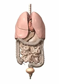
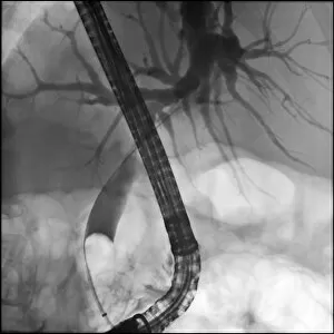
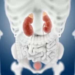
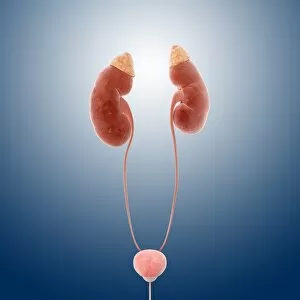
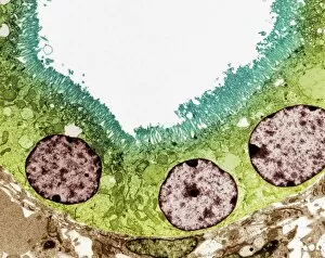
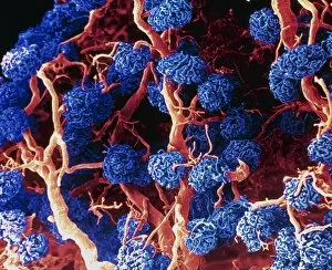
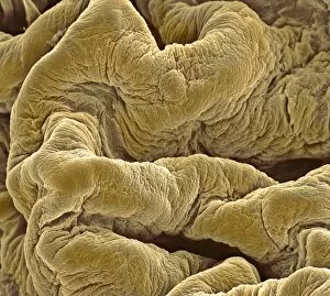
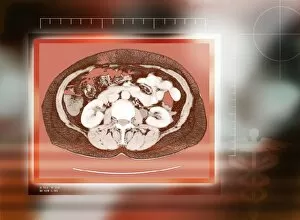
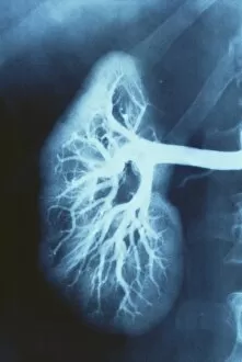
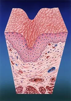
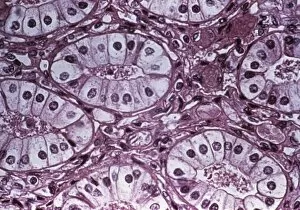
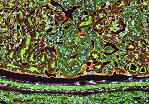
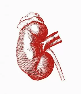
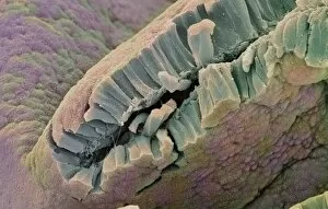
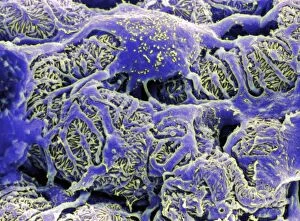
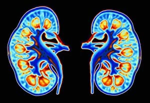
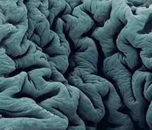
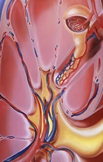
The urinary system is a complex network of organs that play a vital role in maintaining the body's overall health and balance. From the intricate kidney tubules to the colorful MRI scan showcasing the kidneys and liver, this caption explores various aspects of this fascinating system. Starting with a closer look at kidney tubules in section, we delve into their microscopic structure and function. Moving on to an eye-catching color MRI scan of the abdomen, we witness the beauty of these essential organs alongside the liver. Shifting our focus to female anatomy, we encounter artwork depicting both the urinary bladder and a section through it. These illustrations provide insight into its unique composition and how it contributes to waste elimination. Zooming in even further, we explore kidney glomeruli through scanning electron microscopy (SEM). The detailed images reveal their intricate architecture responsible for filtering blood and producing urine. Additionally, SEM captures stunning visuals of kidney blood vessels intertwined within this remarkable filtration process. Further emphasizing their significance, light micrographs showcase multiple kidney glomeruli working harmoniously together. This glimpse highlights their collective effort towards maintaining fluid balance within our bodies. Concluding with captivating artwork portraying kidneys' overall anatomy, we gain a comprehensive understanding of these crucial organs' location and structure. Through this diverse collection of visuals ranging from scientific imaging techniques to artistic representations, one can truly appreciate the complexity and importance of our urinary system.