
Ventricle Collection (#5)
The ventricle, a vital component of the human heart, is beautifully depicted in this collection of artwork and medical imagery


For sale as Licensed Images
Choose your image, Select your licence and Download the media
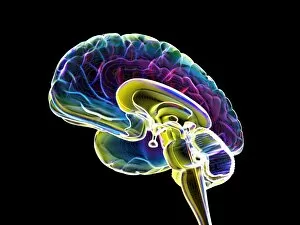
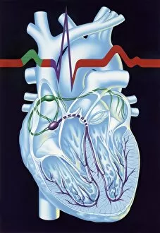
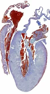
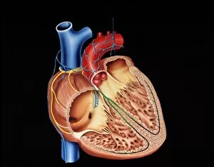
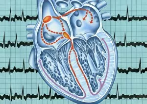
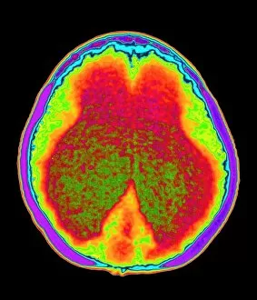
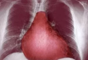
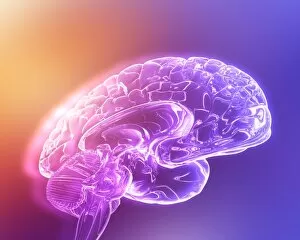
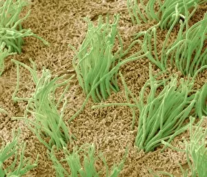
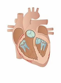
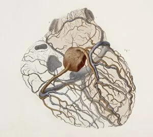
The ventricle, a vital component of the human heart, is beautifully depicted in this collection of artwork and medical imagery. In a late 19th-century French wood engraving, we are presented with a cross-section of the heart, showcasing the intricate network of chambers and vessels that make up this remarkable organ. Moving beyond the surface level, an artwork depicting human heart anatomy provides us with a detailed understanding of how blood flows through the ventricles to ensure proper circulation throughout our bodies. Not limited to just cardiac illustrations, we delve into other fascinating aspects of human biology. An artwork showcasing basal ganglia captures our attention as we explore its role in motor control and cognition. A diagram featuring the heart, lungs, and windpipe reminds us that these organs work in harmony to sustain life-giving oxygenation. Shifting gears towards neuroscience, we encounter captivating images revealing brain structures at different scales. A scanning electron microscope (SEM) image showcases the intricacies of brain surface patterns while an MRI scan unveils both adult and child brains - emphasizing how these structures evolve over time. Returning to matters of cardiovascular health, another cross-section image highlights not only the ventricle but also emphasizes its importance within a healthy heart. This serves as a reminder that maintaining optimal cardiac function is crucial for overall well-being. This diverse collection offers glimpses into various facets of human anatomy - from intricate engravings capturing historical knowledge about hearts to contemporary imaging techniques providing insights into brain development and cardiovascular health. The ventricle takes center stage throughout this compilation as it plays an essential role in sustaining life's most precious resource: our own beating hearts.