
Vertebrae Collection (#2)
"Unlocking the Secrets of Vertebrae: Exploring Chakras and the Nervous System" Delve into the intricate world of vertebrae


For sale as Licensed Images
Choose your image, Select your licence and Download the media
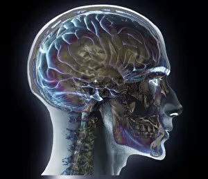
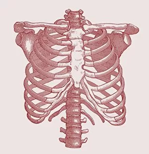
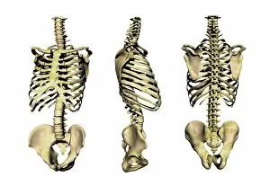
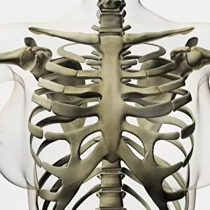
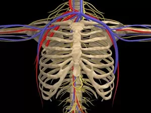
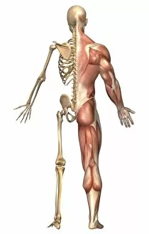
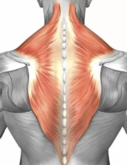
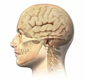
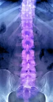
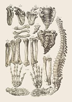
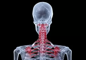
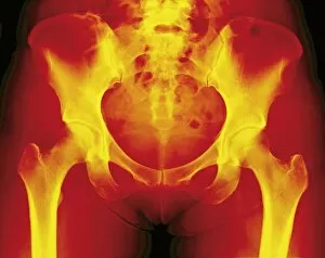
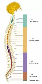
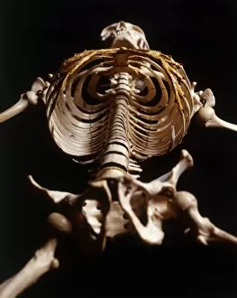
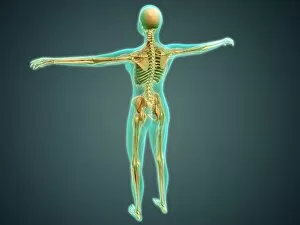
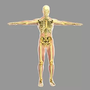
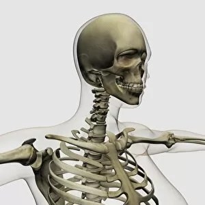
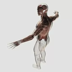
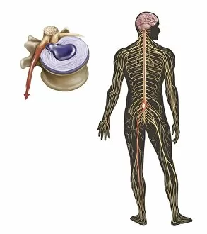
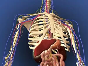
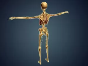
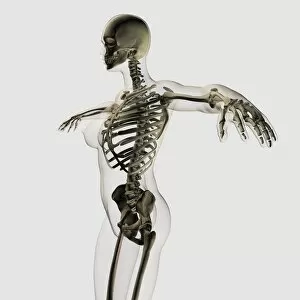
"Unlocking the Secrets of Vertebrae: Exploring Chakras and the Nervous System" Delve into the intricate world of vertebrae, where chakras and the nervous system intertwine like a beautifully orchestrated symphony. A diagram showcasing the human brain and spinal column reveals the remarkable connection between our thoughts and physical well-being. With a camera in hand, capture an X-ray image that unveils both the fragility and strength of our vertebral structure. The upper body skeleton depicted in computer artwork serves as a reminder of how these bones provide support for every movement we make. Observe a normal neck through an X-ray lens, appreciating its graceful alignment that allows us to turn our heads with ease. An X-ray image displaying a normal spine showcases its incredible flexibility while maintaining stability throughout our lives. Through captivating artwork, explore the complexity of human arm musculature intertwined with vertebrae – truly a marvel of nature's design (F007 / 1810). Even animals possess this fascinating skeletal structure; admire a dog skeleton captured in detailed artwork (F006 / 2267). Witness spine repair in action as workers diligently restore damaged vertebrae – highlighting humanity's dedication to healing (F007 / 9907). Plate 19 from Mantell's Geology of Sussex offers insight into how geological forces have shaped even our own vertebral history. A colored X-ray provides a side view glimpse into the delicate intricacies within one's neck – reminding us to cherish this vital part of ourselves. Sometimes, challenges arise such as slipped discs - but modern medicine continues to innovate ways to heal and alleviate discomfort. Intriguingly complex yet undeniably essential, vertebrae serve as both guardians protecting our nervous system and conduits connecting mind and body.