
Vertebral Column Collection (#4)
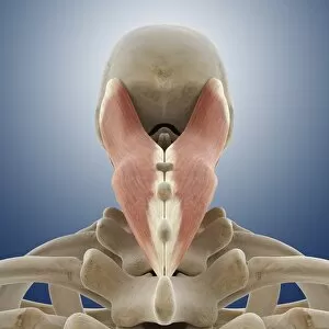
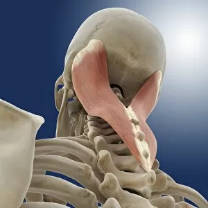
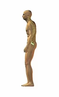
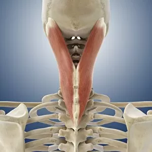
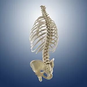
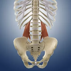
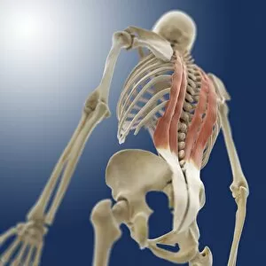
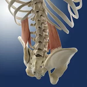
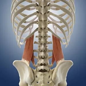
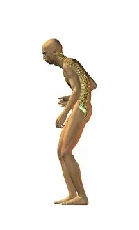
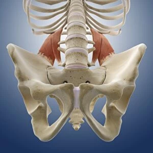
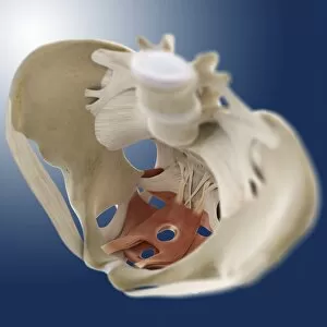
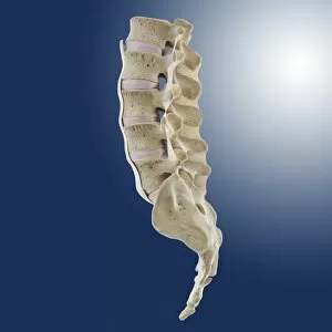
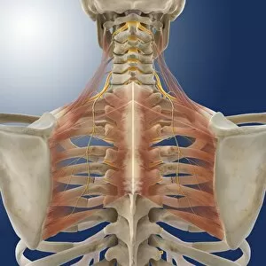
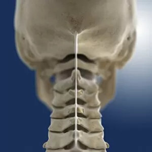
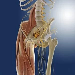
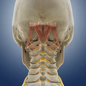
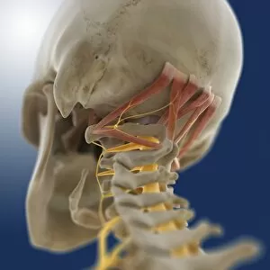
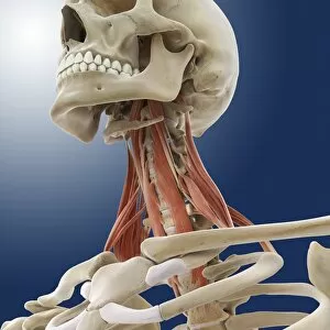
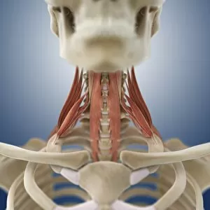
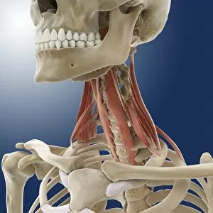
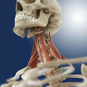
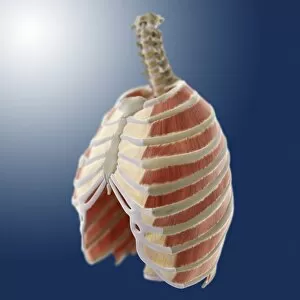
The vertebral column, also known as the backbone or spine, is a crucial structure that provides support and protection to the human body


For sale as Licensed Images
Choose your image, Select your licence and Download the media
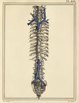
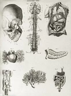
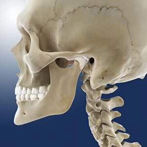
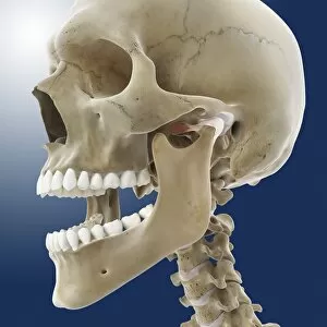
The vertebral column, also known as the backbone or spine, is a crucial structure that provides support and protection to the human body. In a diagram of the human spine from a side view, we can see its intricate arrangement of vertebrae stacked on top of each other like building blocks. This 3D rendering reminds us of the remarkable similarity between our own spinal structure and that of an Ankylosaurus dinosaur skeleton. An X-ray image showcases a normal spine, highlighting its flexibility and alignment. The conceptual image featuring a human skull and spinal cord emphasizes the vital connection between these two essential components. Meanwhile, another 3D rendering displays the imposing skeletal frame of a Tyrannosaurus Rex dinosaur, reminding us that even ancient creatures had their own version of this fundamental anatomical feature. Bartholomeo Eustachi's book "The Science of Human Anatomy" serves as an invaluable resource for understanding the intricacies of the vertebral column. It delves into its composition, function, and importance in maintaining overall bodily health. In contrast to scientific illustrations and historical texts lies an artwork depicting workers repairing a damaged spine - F007 / 9907 - symbolizing medical advancements in treating spinal conditions such as slipped intervertebral discs. Another artistic representation by Santiago Ramon y Cajal portrays colorful histological nerves within the vertebral column; his work earned him recognition with a Nobel Prize in Medicine in 1906. Lithographs showcasing the spinal cord and spinal nerves further emphasize their significance in transmitting signals throughout our body. On a different note, Egon Schiele's painting "Nude with Blue Stockings" captures both vulnerability and grace while bending forward – reminding us how every movement involves coordination from our central axis. Lastly, we witness medicine's progress through time with an image illustrating reduction techniques for dislocated spines where patients are secured onto wooden boards during treatment – highlighting how far we have come in providing care and support for spinal injuries.