
Veterinary Science Collection
"Exploring the Fascinating World of Veterinary Science: From X-rays to Artwork" Step into the captivating realm of veterinary science


For sale as Licensed Images
Choose your image, Select your licence and Download the media
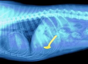
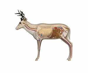
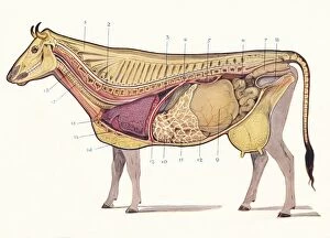
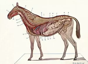
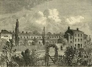
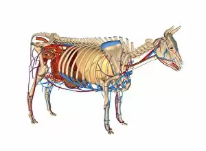
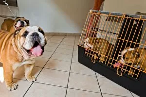
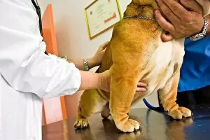
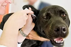
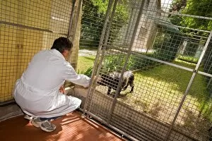
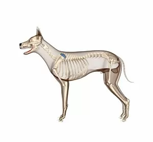
"Exploring the Fascinating World of Veterinary Science: From X-rays to Artwork" Step into the captivating realm of veterinary science, where every day brings new challenges and discoveries. In this intriguing field, even a simple spoon swallowed by a mischievous dog can become an intricate puzzle to solve. Through cutting-edge technology like X-rays, veterinarians unravel mysteries hidden within our beloved animal companions. Delve deeper into the wonders as we explore dog anatomy through stunning artwork. Witness the incredible complexity of their bodies, from muscles and tendons to organs responsible for digestion and circulation. Transport yourself back in time with historical images such as "The Animals Institute-A Hospital for Horses, Dogs, Cats, " established in 1888 by an unknown creator. Travel across borders to Calonne in northern France during the tumultuous years between 1914 and 1918. Behold a veterinary station dedicated solely to horses, showcasing humanity's unwavering commitment to caring for these noble creatures amidst chaos. Marvel at another remarkable image depicting a section of a cow's abdomen with its precious fetus nestled inside – a testament to both life's miracles and veterinary expertise. Explore further as you witness median sections revealing organs vital for circulation, respiration, and more. Unlocking age secrets becomes possible through examining horse teeth – each line etched on their molars tells tales of wisdom gained over time. Delight in studying intricate foot structures that enable these majestic animals' graceful movements. Finally, immerse yourself in RE Holding's breathtaking artwork capturing superficial muscles and tendons adorning horses' powerful frames. A vertical section reveals hidden intricacies beneath their sleek exteriors – truly awe-inspiring. From ancient times when The Royal Agricultural College stood tall in Cirencester until today's modern advancements; veterinary science continues evolving alongside our understanding of animal health care, and is an ever-unfolding journey filled with compassion-driven professionals committed to ensuring the well-being of our cherished animal companions.