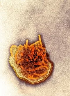
Viral Collection (#10)

"Unveiling the Intricate World of Viruses: From Coronavirus to Norovirus, Hepatitis C to Influenza" In this captivating illustration

Background imageViral Collection: Rabies virus particle, artwork C016 / 9158

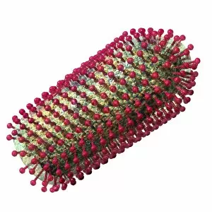
Rabies virus particle, artwork C016 / 9158
Rabies virus particle, computer artwork. This bullet-shaped particle consists of an RNA (ribonucleic acid) genome enclosed in protein capsid

Background imageViral Collection: Measles virus particle, artwork C016 / 9329

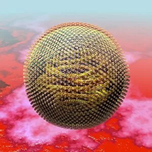
Measles virus particle, artwork C016 / 9329
Measles virus particle, computer artwork. This virus, from the Morbillivirus group of viruses, consists of an RNA (ribonucleic acid)

Background imageViral Collection: Rabies virus particle, artwork C016 / 9157

Rabies virus particle, artwork C016 / 9157
Rabies virus particle, computer artwork. This bullet-shaped particle consists of an RNA (ribonucleic acid) genome enclosed in protein capsid

Background imageViral Collection: Lassa virus particles, TEM C016 / 9409

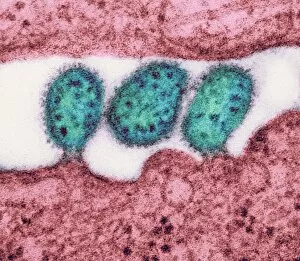
Lassa virus particles, TEM C016 / 9409
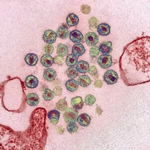
Lassa virus particles. Transmission electron micrograph (TEM) of Lassa virus particles (virions, blue) amongst cell debris. This Arenavirus is the cause of Lassa fever

Background imageViral Collection: Human adenovirus 36, artwork C016 / 8966

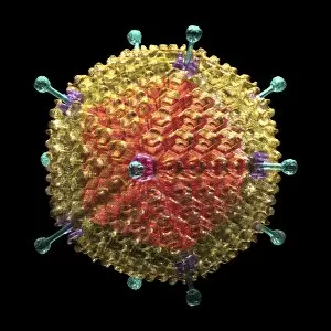
Human adenovirus 36, artwork C016 / 8966
Human adenovirus 36. Computer artwork of human adenovirus 36 (HAdV-36), showing the surface structure of the viruss outer protein coat (capsid)

Background imageViral Collection: Acute haemorrhagic conjunctivitis, TEM C016 / 9387

Acute haemorrhagic conjunctivitis, TEM C016 / 9387
Acute haemorrhagic conjunctivitis. Transmission electron micrograph (TEM) of a sample of tissue from the conjunctiva of a human eye that is infected by enterovirus particles (purple)

Background imageViral Collection: Eastern equine encephalitis, TEM C016 / 9390

Eastern equine encephalitis, TEM C016 / 9390
Eastern equine encephalitis. Transmission electron micrograph (TEM) of a section through tissue from a human central nervous system (CNS)

Background imageViral Collection: Colorado tick fever virus infection, TEM C016 / 9384

Colorado tick fever virus infection, TEM C016 / 9384
Colorado tick fever virus infection. Transmission electron micrograph (TEM) of human tissue infected by Colorado tick fever virus (CTFV) particles (virions, dark circles)

Background imageViral Collection: Swine flu virus particles, TEM C016 / 9399

Swine flu virus particles, TEM C016 / 9399
Swine flu virus particles. Transmission electron micrograph (TEM) of a section through H3N2 influenza A virus particles (virions)

Background imageViral Collection: Rabies virus particles artwork C016 / 9159

Rabies virus particles artwork C016 / 9159
Rabies virus particles, computer artwork. These bullet-shaped particles consist of an RNA (ribonucleic acid) genome enclosed in protein capsid

Background imageViral Collection: Swine flu virus particles, TEM C016 / 9406

Swine flu virus particles, TEM C016 / 9406
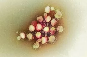
Influenza virus particles. Transmission electron micrograph (TEM) of influenza (flu) virus particles (virions). Each virion consists of ribonucleic acid (RNA, dark patches)

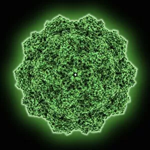
Background imageViral Collection: Adeno-associated virus C016 / 8959

Adeno-associated virus C016 / 8959
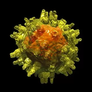
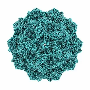
Adeno-associated virus, computer artwork. Adeno- associated viruses (aVs) are the smallest known viruses to infect humans. They do not cause diseases, and only provoke a mild immune response

Background imageViral Collection: Rabies virus particle, artwork C016 / 9155

Rabies virus particle, artwork C016 / 9155
Rabies virus particle, computer artwork. This bullet-shaped particle consists of an RNA (ribonucleic acid) genome enclosed in protein capsid

Background imageViral Collection: HIV virus particles, TEM C016 / 9404

HIV virus particles, TEM C016 / 9404
HIV virus particles. Transmission electron micrograph (TEM) of a section through human immunodeficiency virus (HIV) particles (virions, round)

Background imageViral Collection: HIV virus particles, TEM C016 / 9405

HIV virus particles, TEM C016 / 9405
HIV virus particles. Transmission electron micrograph (TEM) of a section through human immunodeficiency virus (HIV) particles (virions, round)

Background imageViral Collection: Swine flu virus particles, TEM C016 / 9400

Swine flu virus particles, TEM C016 / 9400
Swine flu virus particles. Transmission electron micrograph (TEM) of a section through H3N2 influenza A virus particles (virions)

Background imageViral Collection: Adeno-associated virus C016 / 8958

Adeno-associated virus C016 / 8958
Adeno-associated virus, computer artwork. Adeno- associated viruses (aVs) are the smallest known viruses to infect humans. They do not cause diseases, and only provoke a mild immune response

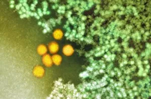
Background imageViral Collection: Lassa virus particles, TEM C016 / 9408

Lassa virus particles, TEM C016 / 9408
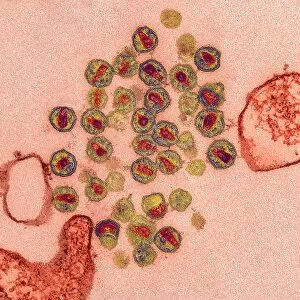
Lassa virus particles. Transmission electron micrograph (TEM) of Lassa virus particles (virions, green) amongst cell debris. This Arenavirus is the cause of Lassa fever

Background imageViral Collection: Rabies virus particle, artwork C016 / 9156

Rabies virus particle, artwork C016 / 9156
Rabies virus particle, computer artwork. This bullet-shaped particle consists of an RNA (ribonucleic acid) genome enclosed in protein capsid

Background imageViral Collection: Ganjam virus infection, TEM C016 / 9396

Ganjam virus infection, TEM C016 / 9396
Ganjam virus infection. Transmission electron micrograph (TEM) of a section through human tissue infected with ganjam virus particles (virions, blue and yellow)
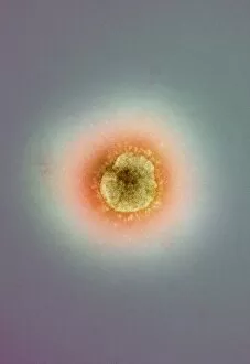
Background imageViral Collection: Swine flu virus particles, TEM C016 / 9407

Swine flu virus particles, TEM C016 / 9407
Influenza virus particles. Transmission electron micrograph (TEM) of influenza (flu) virus particles (virions). Each virion consists of ribonucleic acid (RNA, dark patches)

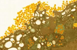
Background imageViral Collection: Schmallenberg virus particles, TEM

Schmallenberg virus particles, TEM
Schmallenberg virus. Transmission electron micrograph (TEM) of virus particles (virions, yellow) of the Schmallenberg virus

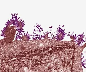
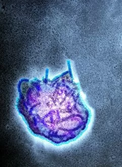
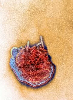
Background imageViral Collection: Flu virus, TEM

Flu virus, TEM
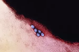
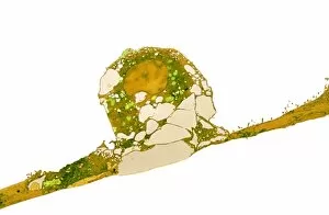
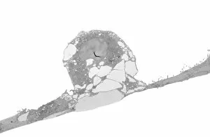
Flu virus. Transmission electron micrograph (TEM) of a section through influenza (flu) virus particles (virions, purple) budding from a host cell. This is the final stage in viral replication

Background imageViral Collection: Bird flu, conceptual artwork

Bird flu, conceptual artwork
Bird flu. Conceptual artwork of a chicken and flying syringes, representing measures taken against the spread of avian influenza (bird flu)

Background imageViral Collection: Genomic HIV-RNA duplex

Genomic HIV-RNA duplex, molecular model. This structure shows the dimerization initiation site of genomic HIV-1 with RNA (ribonucleic acid)

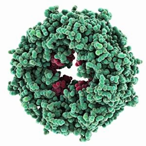
Background imageViral Collection: Ebola matrix protein molecule

Ebola matrix protein molecule
Ebola matrix protein. Molecular model of the Ebola virus matrix protein VP40 (green) bound to RNA (ribonucleic acid, red)

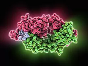
Background imageViral Collection: HIV antibody and glycoprotein complex

HIV antibody and glycoprotein complex. Molecular model of the anti-human immunodeficiency virus type 1 (anti-HIV-1) antibody 2F5 in complex with the gp41 envelope glycoprotein of HIV-1

Background imageViral Collection: MERS coronavirus, TEM C015 / 1772

MERS coronavirus, TEM C015 / 1772
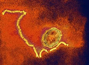
MERS coronavirus, coloured transmission electron micrograph (TEM). This virus (originally novel coronavirus 2012) has been named Middle East respiratory syndrome (MERS) coronavirus

Background imageViral Collection: MERS coronavirus, TEM C015 / 1774

MERS coronavirus, TEM C015 / 1774
MERS coronavirus, coloured transmission electron micrograph (TEM). This virus (originally novel coronavirus 2012) has been named Middle East respiratory syndrome (MERS) coronavirus

Background imageViral Collection: MERS coronavirus, TEM C015 / 1776

MERS coronavirus, TEM C015 / 1776
MERS coronavirus, coloured transmission electron micrograph (TEM). This virus (originally novel coronavirus 2012) has been named Middle East respiratory syndrome (MERS) coronavirus

Background imageViral Collection: MERS coronavirus, TEM C015 / 1773

MERS coronavirus, TEM C015 / 1773
MERS coronavirus, coloured transmission electron micrograph (TEM). This virus (originally novel coronavirus 2012) has been named Middle East respiratory syndrome (MERS) coronavirus

Background imageViral Collection: Avian influenza virus H7N9, TEM C016 / 6294

Avian influenza virus H7N9, TEM C016 / 6294
Avian influenza virus, type A strain H7N9, coloured transmission electron micrograph (TEM). This virus first emerged in the human population in China, in March 2013

Background imageViral Collection: Avian influenza virus H7N9, TEM C016 / 6293

Avian influenza virus H7N9, TEM C016 / 6293
Avian influenza virus, type A strain H7N9, coloured transmission electron micrograph (TEM). This virus first emerged in the human population in China, in March 2013

Background imageViral Collection: Avian influenza virus H7N9, TEM C016 / 6292

Avian influenza virus H7N9, TEM C016 / 6292
Avian influenza virus, type A strain H7N9, coloured transmission electron micrograph (TEM). This virus first emerged in the human population in China, in March 2013

Background imageViral Collection: Canine parvovirus capsid C015 / 8460

Canine parvovirus capsid C015 / 8460
Canine parvovirus capsid, molecular model. Canine parvovirus type 2 (CPV2) was first recognized in 1978 and spread worldwide in less than two years

Background imageViral Collection: Measles virus particle, TEM C015 / 7160

Measles virus particle, TEM C015 / 7160
Measles virus particle, coloured transmission electron micrograph (TEM). This virus, from the Morbillivirus group of viruses, consists of an RNA (ribonucleic acid)

Background imageViral Collection: Measles virus particle, TEM C015 / 7161

Measles virus particle, TEM C015 / 7161
Measles virus particle, coloured transmission electron micrograph (TEM). This virus, from the Morbillivirus group of viruses, consists of an RNA (ribonucleic acid)

Background imageViral Collection: MERS coronavirus, TEM C015 / 7155

MERS coronavirus, TEM C015 / 7155
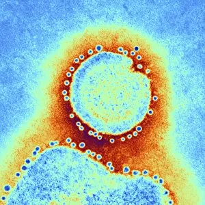
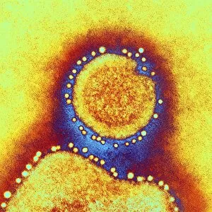
MERS coronavirus. Coloured transmission electron micrograph (TEM) of MERS coronavirus particles (orange) budding from a host cell (brown)

Background imageViral Collection: Bird flu research C015 / 8803

Bird flu research C015 / 8803
Bird flu research. Researcher harvesting influenza type a H7N9 viruses from cultures grown in eggs. This virus first emerged in the human population in China, in March 2013

Background imageViral Collection: MERS coronavirus, TEM C015 / 7157

MERS coronavirus, TEM C015 / 7157
MERS coronavirus. Coloured transmission electron micrograph (TEM) of MERS coronavirus particles (small green dots) budding from a host cell (brown)

Background imageViral Collection: MERS coronavirus, TEM C015 / 7158

MERS coronavirus, TEM C015 / 7158
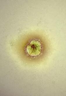
MERS coronavirus. Transmission electron micrograph (TEM) of MERS coronavirus particles (small dots) budding from a host cell (centre)

Background imageViral Collection: Canine parvovirus capsid C015 / 8459

Canine parvovirus capsid C015 / 8459
Canine parvovirus capsid, molecular model. Canine parvovirus type 2 (CPV2) was first recognized in 1978 and spread worldwide in less than two years

Background imageViral Collection: Avian influenza virus, TEM C015 / 8800

Avian influenza virus, TEM C015 / 8800
Avian influenza virus, type A strain H7N9, coloured transmission electron micrograph (TEM). This virus first emerged in the human population in China, in March 2013

Background imageViral Collection: Measles virus particle, TEM C015 / 7162

Measles virus particle, TEM C015 / 7162
Measles virus particle, coloured transmission electron micrograph (TEM). This virus, from the Morbillivirus group of viruses

Background imageViral Collection: Bird flu diagnostic kit C015 / 8805

Bird flu diagnostic kit C015 / 8805
Bird flu diagnostic kit. Reagents for diagnosing influenza type a H7N9 infection in humans. This virus first emerged in the human population in China, in March 2013

Background imageViral Collection: Measles virus particle, TEM C015 / 7159

Measles virus particle, TEM C015 / 7159
Measles virus particle, coloured transmission electron micrograph (TEM). This virus, from the Morbillivirus group of viruses, consists of an RNA (ribonucleic acid)

Background imageViral Collection: Avian influenza virus, TEM C015 / 8799

Avian influenza virus, TEM C015 / 8799
Avian influenza virus, type A strain H7N9, coloured transmission electron micrograph (TEM). This virus first emerged in the human population in China, in March 2013

Background imageViral Collection: Bird flu research C015 / 8804

Bird flu research C015 / 8804
Bird flu research. Researcher measuring harvested influenza type a H7N9 viruses grown in the lab. This virus first emerged in the human population in China, in March 2013


For sale as Licensed Images

Choose your image, Select your licence and Download the media

"Unveiling the Intricate World of Viruses: From Coronavirus to Norovirus, Hepatitis C to Influenza" In this captivating illustration, we delve into the microscopic realm of viruses that have left an indelible mark on our lives. Firstly, we encounter a mesmerizing depiction of the Coronavirus Structure Illustration. Its intricate spikes and spherical shape remind us of its global impact and ongoing battle against humanity. Moving forward, we witness Norovirus particles through a powerful TEM image. These tiny entities are notorious for causing stomach upsets and outbreaks in various settings. Next, our attention is drawn towards a molecular model showcasing the Hepatitis C virus enzyme. This visual representation highlights the complexity behind this viral infection that affects millions worldwide. As we explore further, an artistic rendering presents Flu virus particle artwork F008/3245. It serves as a reminder of how influenza can swiftly spread across populations during seasonal outbreaks. The dangers posed by infections transmitted through sneezing are vividly depicted in artwork C013/5949. The visualization captures the moment when pathogens disperse into the air with every forceful expulsion from an infected individual's respiratory system. Returning to coronavirus territory, another TEM image showcases Coronavirus particles in all their glory. Their distinctive crown-like appearance reinforces their notoriety as agents responsible for severe respiratory illnesses like SARS and MERS. Shifting gears slightly, an artistic portrayal introduces us to Adenovirus - known for causing respiratory infections but also harnessed as vectors in gene therapy research due to their unique properties. Our journey continues with yet another TEM image revealing Influenza virus particles - these minuscule culprits have been responsible for numerous pandemics throughout history and continue to pose significant health threats today. Stepping away momentarily from respiratory viruses, we observe Herpes virus replicating within cells under computer-generated artwork. This glimpse into its life cycle reminds us of the persistence and challenges associated with managing this widespread infection.