
Visual Sense Collection (page 2)
"Unlocking the Wonders of Visual Sense: Exploring the Intricacies of Our Eyes" Our eyes, like intricate masterpieces, hold within them a world of visual wonders


For sale as Licensed Images
Choose your image, Select your licence and Download the media
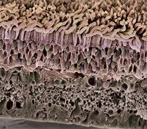
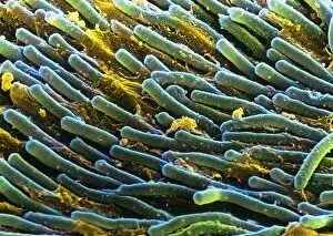
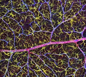
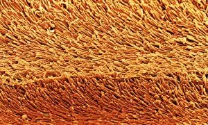
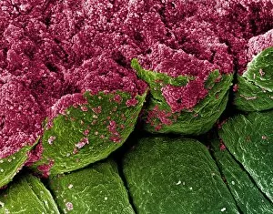
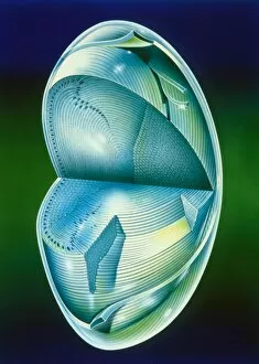
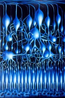
"Unlocking the Wonders of Visual Sense: Exploring the Intricacies of Our Eyes" Our eyes, like intricate masterpieces, hold within them a world of visual wonders. The iris, akin to a delicate flower's petals, reveals its captivating hues and patterns that make each individual unique. Through the lens of a scanning electron microscope (SEM), we delve into the mesmerizing details of this remarkable feature. As we journey deeper into our visual sense, contact lenses emerge as modern marvels that enhance our perception. They seamlessly blend with our irises, offering clarity and comfort while allowing us to see the world in all its vibrant glory. The retina takes center stage in this symphony of sight – an intricately woven tapestry capturing light and transforming it into vivid images. Its false-color SEM unveils the central fovea, where vision is sharpest and most precise; a testament to nature's precision craftsmanship. Delving further into this microscopic realm, we encounter the ciliary body – an unsung hero responsible for adjusting our eye's focus effortlessly. Revealed by SEM imaging techniques, its intricate structures showcase nature's ingenuity at work. Traversing through retinal blood vessels captured by SEM imagery unravels their complex network nourishing every corner of our visual canvas. These lifelines remind us how interconnected every aspect of our visual sense truly is. The eye lens emerges as another extraordinary component under scrutiny by SEM technology - revealing its layered architecture designed meticulously for optimal refraction and focusing power. It serves as a gateway between worlds - bending light rays to create clear visions before they reach our precious retina. Within this masterpiece lies rods and cones - tiny warriors guarding against darkness or illuminating colors with finesse unmatched elsewhere in nature’s palette. Their artistic portrayal showcases their vital role in shaping what we perceive around us. Lastly but not leastly comes eye color – an enchanting kaleidoscope reflecting both genetics and individuality.