
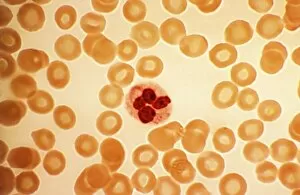
White Blood Cell Collection
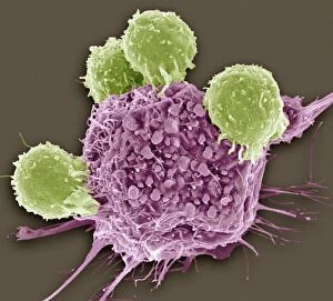
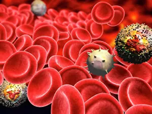
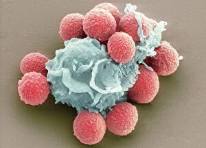
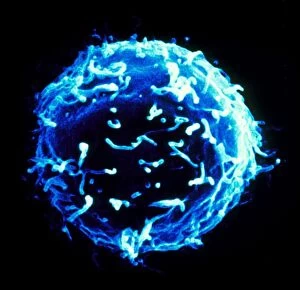
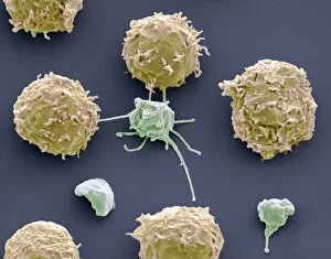
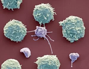
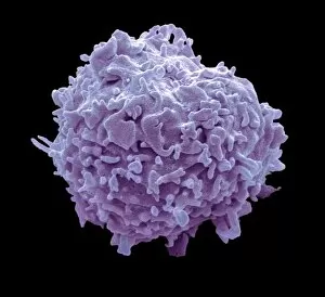
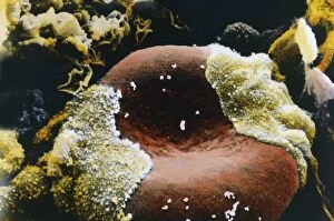
"Guardians of Health: Exploring the Mighty White Blood Cell" T lymphocytes and cancer cells: Unveiling the battle within, as T lymphocytes wage war against cancer cells
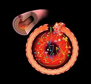


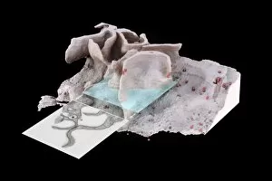

Digital cross section illustration wound below human skin showing red and white blood cells and yell

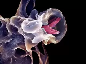
Digital cross section illustration of ciliate cell showing rhinovirus and antobodies in nasal cavity


For sale as Licensed Images
Choose your image, Select your licence and Download the media
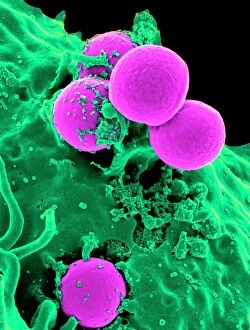
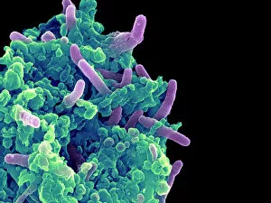
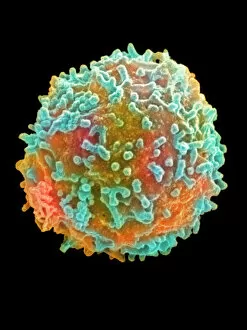
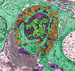
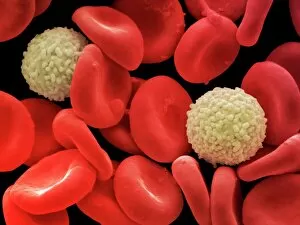
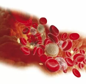
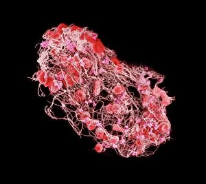
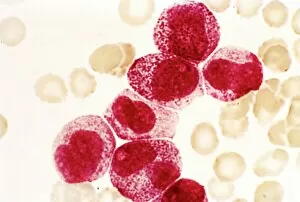
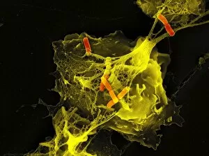
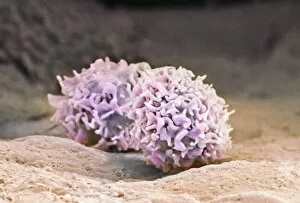
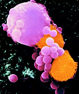
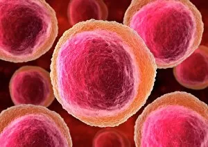
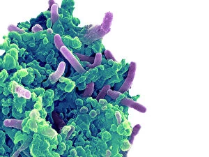
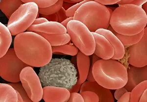
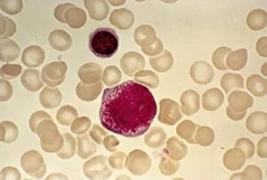
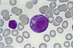
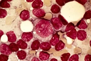
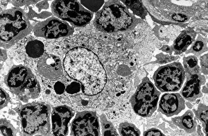
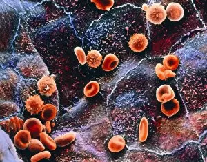
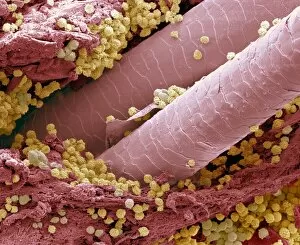
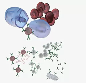
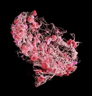
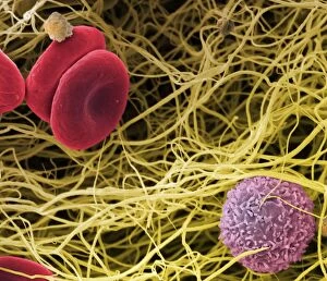
"Guardians of Health: Exploring the Mighty White Blood Cell" T lymphocytes and cancer cells: Unveiling the battle within, as T lymphocytes wage war against cancer cells, SEM C001 / 1679. Neutrophil engulfing MRSA: Witness the incredible defense mechanism as a neutrophil engulfs MRSA bacteria, SEM C018 / 8596. Dendritic cells artwork: Unlocking the secrets of immune response with stunning artwork depicting dendritic cells at work. TEM of human white blood cell bearing HLA antigen: Peering into the intricate world of immunity through a transmission electron microscope image showcasing a white blood cell displaying HLA antigens. Blood cells in harmony: A mesmerizing glimpse into our life force - an ensemble of diverse blood cells working together for our well-being. Coloured SEM of a white blood cell (lymphocyte): Dive deep into the vibrant realm of lymphocytes captured in this captivating colored scanning electron microscope image. Basophil white blood cell: Discovering the lesser-known heroes among us - basophilic white blood cells that play crucial roles in allergic reactions and inflammation. Bacteria infecting a macrophage, SEM: Witness how macrophages confront invading bacteria head-on under high-resolution scanning electron microscopy imagery. Blood coagulation cascade artwork: Unraveling the intricacies behind clot formation with an artistic representation capturing every step in this vital process, artwork C016 / 9873 Red and white blood cells, SEM : Marvel at nature's palette as red and white blood cells come alive under scanning electron microscopy - an exquisite symphony within us all. Blood clot, SEM C016 / 9747 : Delving into hemostasis marvels with a striking scanning electron micrograph revealing intricate details within a formed clot structure. Dohle bodies in blood cell, micrograph.