Hibiscus seeds

![]()
Wall Art and Photo Gifts from Mary Evans Picture Library
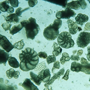
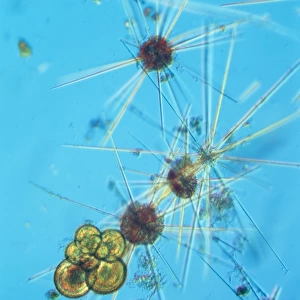
Hibiscus seeds
An illustration of three hibiscus seeds as seen through a microscope, from page 497 of Flora, overo Cultura di Fiori (1638) by Battista Giovanni Ferrari (1582-1655)
Mary Evans Picture Library makes available wonderful images created for people to enjoy over the centuries
Media ID 8601515
© Mary Evans Picture Library 2015 - https://copyrighthub.org/s0/hub1/creation/maryevans/MaryEvansPictureID/10705187
17th Century Eudicot Eurosid Ferrari Flora Hibiscus Malvaceae Malvales Malvidae Microscopic Publication Rosid Seed Angiospermae Dicot Dicotyledon Magnoliophyta
EDITORS COMMENTS
This illustration showcases three hibiscus seeds as seen through the lens of a microscope, a fascinating glimpse into the intricacies of nature from the 17th century. The illustration is taken from Flora, overo Cultura di Fiori (Flowers, or The Cultivation of Flowers), a botanical publication by Battista Giovanni Ferrari, an Italian botanist and artist born in 1582. Ferrari's extensive work on botany and horticulture spanned over seven decades, making significant contributions to the scientific community during the Renaissance period. The illustration, found on page 497 of the publication, provides a detailed view of the hibiscus seeds, which belong to the Malvaceae family, a large and diverse group of flowering plants commonly known as mallow or hibiscus. As angiosperms, dicotyledons, or eudicots, hibiscus plants are characterized by their two seed leaves and net-like veins. The seeds themselves are enclosed within a fruit called a capsule, which splits open to release the seeds upon maturity. The illustration's historical significance lies in its meticulous attention to detail and its role in advancing the scientific understanding of plant morphology during the 17th century. Ferrari's work was instrumental in documenting the botanical world and expanding European knowledge of exotic plants. This image of hibiscus seeds, viewed through the microscope, offers a glimpse into the intricacies of the natural world that were only beginning to be explored during this time. The illustration is a testament to Ferrari's artistic skill and his deep understanding of the botanical world. It is a reminder of the importance of documenting the natural world and the role that illustrations play in advancing scientific knowledge. This image continues to inspire curiosity and wonder, inviting us to explore the intricacies of the natural world and the beauty of the plants that surround us.
MADE IN THE USA
Safe Shipping with 30 Day Money Back Guarantee
FREE PERSONALISATION*
We are proud to offer a range of customisation features including Personalised Captions, Color Filters and Picture Zoom Tools
SECURE PAYMENTS
We happily accept a wide range of payment options so you can pay for the things you need in the way that is most convenient for you
* Options may vary by product and licensing agreement. Zoomed Pictures can be adjusted in the Cart.