Home > Mary Evans Prints Online > New Images August 2021
Anatomy of the human ear and eye

![]()
Wall Art and Photo Gifts from Mary Evans Picture Library
Anatomy of the human ear and eye
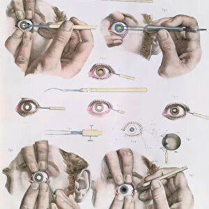
Anatomy of the human ear and eye. Lithograph from Lorenz Okens Universal Natural History, Allgemeine Naturgeschichte fur alle Stande, Stuttgart, 1839
Mary Evans Picture Library makes available wonderful images created for people to enjoy over the centuries
Media ID 23196814
© Florilegius/Mary Evans
Alle Allgemeine Eyeball Hearing Human Lens Lorenz Naturgeschichte Nerves Oken Sense Sight Stände Universal Retina
EDITORS COMMENTS
This stunning lithograph, titled "Anatomy of the Human Ear and Eye," is an exquisite illustration from Lorenz Oken's comprehensive work, "Allgemeine Naturgeschichte fur alle Stände," published in Stuttgart, Germany, in 1839. The image provides an intricate and detailed view of the anatomy of two of the most complex and essential organs in the human body: the ear and the eye. The ear, depicted on the left side of the print, is shown in cross-section, revealing its intricate labyrinth of structures. The outer ear collects sound waves and channels them into the ear canal, leading to the eardrum. The middle ear contains the ossicles, tiny bones that amplify sound, and the inner ear houses the cochlea, where sound is converted into electrical signals that are sent to the brain. Nerves and various structures, including the vestibular system for balance, are also illustrated. The eye, on the right side of the print, is shown in a similar manner, with its various components meticulously labeled. The eyeball is divided into the anterior and posterior segments. The anterior segment includes the cornea, iris, and lens, which focus light onto the retina. The posterior segment consists of the choroid, retina, and optic nerve, which transmit the visual information to the brain. This print, created by the skilled hands of a master lithographer, offers a fascinating glimpse into the inner workings of the human senses of sight and hearing. It is a testament to the scientific curiosity and artistic mastery of the 19th century, and a reminder of the enduring importance of understanding the intricacies of our own bodies.
MADE IN THE USA
Safe Shipping with 30 Day Money Back Guarantee
FREE PERSONALISATION*
We are proud to offer a range of customisation features including Personalised Captions, Color Filters and Picture Zoom Tools
SECURE PAYMENTS
We happily accept a wide range of payment options so you can pay for the things you need in the way that is most convenient for you
* Options may vary by product and licensing agreement. Zoomed Pictures can be adjusted in the Cart.