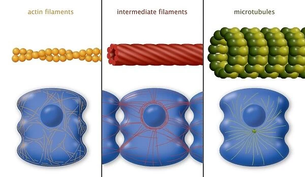
Cytoskeleton components, diagram
![]()

Wall Art and Photo Gifts from Science Photo Library
Cytoskeleton components, diagram
Cytoskeleton components, diagram. The cytoskeleton is the internal support structure of a cell, composed of filaments of various diameters in nanometres (nm). At left, the globular protein actin (orange) forms a microfilament (6nm). At centre, intermediate filaments (red, 10nm) are composed of a family of various proteins. The thickest of the filaments are the microtubules (green, right, 25nm), composed of the protein tubulin. Some of their roles in a cell are shown across bottom. From left to right: cell contraction and movement; cell nucleus and desmosomes (cell-cell adhesions); and a mitotic spindle (below the nucleus)
Science Photo Library features Science and Medical images including photos and illustrations
Media ID 6326909
© ART FOR SCIENCE/SCIENCE PHOTO LIBRARY
Actin Actin Filament Assembly Cell Biology Cell Nucleus Cellular Component Contraction Cytoskeleton Filament Filaments Intermediate Filament Large Macromolecule Medium Microtubule Microtubules Mitosis Molecular Biology Movement Polymer Small Structural Thick Thin Trio Tubulin Type Types Bio Chemistry Biochemical Protein
EDITORS COMMENTS
This print from Science Photo Library showcases the intricate components of the cytoskeleton, which serves as the internal support structure of a cell. The diagram highlights three types of filaments with varying diameters in nanometres, each playing crucial roles within cellular processes. On the left side, we see actin, a globular protein forming microfilaments measuring 6nm in diameter. Actin filaments are involved in cell contraction and movement, enabling cells to change shape and migrate. In the center, intermediate filaments colored red (10nm) consist of a diverse family of proteins. These medium-sized filaments contribute to structural integrity and provide mechanical strength to cells. They are also essential for maintaining cell adhesions called desmosomes and supporting the nucleus. The thickest among these filamentous structures are microtubules depicted on the right side in green (25nm). Composed of tubulin proteins, microtubules have multiple functions including facilitating cell motility by acting as tracks for molecular motors during intracellular transport. Additionally, they form a mitotic spindle below the nucleus during cell division (mitosis), ensuring accurate chromosome segregation. This visually stunning artwork beautifully captures the complexity and diversity present within cellular biology. It emphasizes how these cytoskeletal components work together harmoniously to maintain cellular structure and enable vital biological processes such as movement and division.
MADE IN THE USA
Safe Shipping with 30 Day Money Back Guarantee
FREE PERSONALISATION*
We are proud to offer a range of customisation features including Personalised Captions, Color Filters and Picture Zoom Tools
SECURE PAYMENTS
We happily accept a wide range of payment options so you can pay for the things you need in the way that is most convenient for you
* Options may vary by product and licensing agreement. Zoomed Pictures can be adjusted in the Cart.

