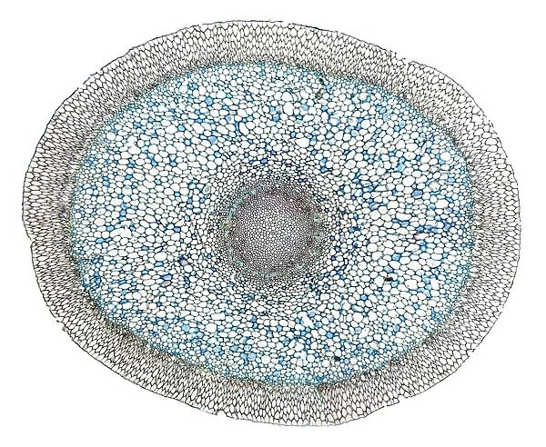
Dendrobium orchid root, light micrograph
![]()

Wall Art and Photo Gifts from Science Photo Library
Dendrobium orchid root, light micrograph
Dendrobium orchid root. Light micrograph of a section through an aerial root from a Dendrobium sp. orchid. The outer tissue (velamen radicum, grey) is composed of hexagonal cells. These are dead cells with lignified walls that contain pores that take up water. The next layer, the exodermis, is a ring of cells with walls of suberin that contain passage cells for water flow. The next layer is the cortex of parenchyma cells (blue). The central core has an outer ring (endodermis) with thick walls (grey) and next a ring of vascular bundles with alternate rings of xylem (black) and phloem (green). Magnification: x13 when printed 10 centimetres wide
Science Photo Library features Science and Medical images including photos and illustrations
Media ID 6353949
© DR KEITH WHEELER/SCIENCE PHOTO LIBRARY
Aerial Root Bundle Cell Biology Cell Wall Cortex Cytological Cytology Endodermis Histological Histology Microscopy Monocot Monocots Monocotyledon Monocotyledons Orchid Parenchyma Phloem Stain Stained Structural Structures Tissue Vascular Bundles Xylem Cells Light Micrograph Light Microscope Section Sectioned
EDITORS COMMENTS
This print showcases the intricate beauty of a Dendrobium orchid root, as seen through a light micrograph. The image reveals the various layers and structures that make up this remarkable botanical specimen. Starting from the outer tissue known as velamen radicum, we can observe hexagonal cells with lignified walls that serve as water-absorbing pores. Moving inward, we encounter the exodermis, which consists of suberin-walled cells responsible for facilitating water flow. The next layer is comprised of parenchyma cells forming the cortex, depicted in a striking blue hue. As we delve deeper into the core, an outer ring called endodermis emerges with its thick grey walls. Surrounding it are vascular bundles arranged in alternating rings of xylem (black) and phloem (green), essential for transporting nutrients throughout the plant. With a magnification level of x13 when printed at 10 centimeters wide, this photograph allows us to appreciate every minute detail within this microscopic world. It serves as a testament to both the structural complexity and delicate elegance found within nature's creations. This stunning image provides valuable insights into botany and cell biology while showcasing the unique characteristics specific to dendrobium orchids. Whether you are an enthusiast or scientist exploring histology or cytological studies, this print offers an awe-inspiring glimpse into one of nature's wonders – capturing not only its scientific significance but also its inherent artistic appeal.
MADE IN THE USA
Safe Shipping with 30 Day Money Back Guarantee
FREE PERSONALISATION*
We are proud to offer a range of customisation features including Personalised Captions, Color Filters and Picture Zoom Tools
SECURE PAYMENTS
We happily accept a wide range of payment options so you can pay for the things you need in the way that is most convenient for you
* Options may vary by product and licensing agreement. Zoomed Pictures can be adjusted in the Cart.

