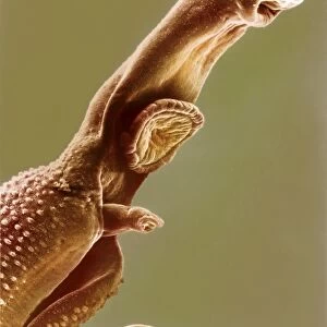
E. coli and Shigella sp. bacteria

![]()
Wall Art and Photo Gifts from Science Photo Library
E. coli and Shigella sp. bacteria
E. coli and Shigella sp. bacteria. Fluorescence confocal light micrograph of Escherichia coli and Shigella sp. bacteria (blue) in human Caco-2 cells (green), a cultured cell line derived from a colorectal carcinoma. The cell nuclei are red. These are motile bacteria that are capable of entering intestinal cells (enteroinvasive) and causing disease. Shigella sp. bacteria cause dysentery, which can vary in severity from a mild attack of diarrhoea to an acute infection. Enteroinvasive E. coli cause severe diarrhoea that can be fatal, especially in the very young or elderly
Science Photo Library features Science and Medical images including photos and illustrations
Media ID 6293047
© STEPHANIE SCHULLER/SCIENCE PHOTO LIBRARY
Bacteria Bacterial Bacteriology Bacterium C Ulture Confocal Light Micrograph Diarrhoea Ecoli Escherichia Coli Fluorescence Fluorescence Light Micrograph Fluorescent Gram Negative Immunofluorescence Immunofluorescent Infected Infecting Infection Intestinal Micro Organisms Microbe Microbes Motile Pathogenic Rods Dysentry Light Micrograph Light Microscope Micro Biology Microbiological Pathogen
EDITORS COMMENTS
This print from Science Photo Library showcases the intricate world of bacteria. In this fluorescence confocal light micrograph, we are presented with a mesmerizing display of Escherichia coli and Shigella sp. bacteria in human Caco-2 cells derived from a colorectal carcinoma. The vibrant blue hue represents these motile bacteria, known for their ability to invade intestinal cells and cause disease. Shigella sp. bacteria specifically induce dysentery, which can range from mild diarrhea to severe infections. On the other hand, enteroinvasive E. coli is responsible for severe and potentially fatal cases of diarrhea, particularly among vulnerable populations such as young children and the elderly. The green coloration signifies the cultured cell line within which these pathogens thrive, while the red highlights emphasize the presence of cell nuclei amidst this microscopic battleground. This image serves as a reminder of both the beauty and danger that exist within our biological landscape. Through advanced imaging techniques like immunofluorescence and confocal microscopy, scientists gain invaluable insights into pathogenic mechanisms at play in infectious diseases. Science Photo Library continues to provide us with awe-inspiring visuals that bridge artistry with scientific discovery - an essential resource for researchers, educators, and anyone fascinated by microbiology's hidden realms. "
MADE IN THE USA
Safe Shipping with 30 Day Money Back Guarantee
FREE PERSONALISATION*
We are proud to offer a range of customisation features including Personalised Captions, Color Filters and Picture Zoom Tools
SECURE PAYMENTS
We happily accept a wide range of payment options so you can pay for the things you need in the way that is most convenient for you
* Options may vary by product and licensing agreement. Zoomed Pictures can be adjusted in the Cart.