Inner ear anatomy, artwork

![]()
Wall Art and Photo Gifts from Science Photo Library
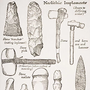
Inner ear anatomy, artwork
Inner ear anatomy. Artwork showing the anatomy of the inner human ear, the organ of hearing and balance. The ear canal (bottom) leads from the pinna (not seen), the visible part of the ear, to the eardrum (tympanic membrane, green), which separates the outer and middle ear. The eardrum transmits sounds, as vibrations, from the air to the bones (ossicles), of the middle ear. These bones, from bottom left to top right, are the malleus (hammer), incus (anvil) and stapes (stirrup). The ossicles join to the inner ear, which consists of fluid-filled passages called the labyrinth. This includes the cochlea (orange semicircle at right), which translates the vibrations into electrical impulses that are carried to the brain by nerves, and the semi-circular canals (red), which are responsible for balance
Science Photo Library features Science and Medical images including photos and illustrations
Media ID 6347911
© FRANCIS LEROY, BIOCOSMOS/SCIENCE PHOTO LIBRARY
Anvil Auditory Nerve Auditory Sense Aural Balance Bones Canals Cochlea Cochlear Ear Canal Ear Drum Hammer Hearing Incus Inner Ear Labyrinth Malleus Middle Ear Ossicle Ossicles Smallest Stapes Stirrup Tympanic Membrane Section Sectioned
EDITORS COMMENTS
This artwork showcases the intricate inner ear anatomy, a remarkable organ responsible for both hearing and balance. Against a striking black background, this illustration delves into the depths of our auditory system. The image begins with the ear canal at the bottom, leading from the unseen pinna (visible part of the ear) to the vibrant green eardrum or tympanic membrane that separates the outer and middle ear. The eardrum plays a crucial role in transmitting sound vibrations from air to three tiny bones known as ossicles: malleus (hammer), incus (anvil), and stapes (stirrup). These delicate bones connect to an awe-inspiring labyrinth within our inner ear, filled with fluid-filled passages. At its core lies the cochlea, depicted as an orange semicircle on the right side of this artwork. This extraordinary structure translates vibrations into electrical impulses that are then carried by nerves to our brain. Additionally, we can observe red semi-circular canals within this labyrinth which contribute significantly to maintaining our sense of balance. This comprehensive representation provides insight into how these interconnected components work harmoniously together. With its meticulous detail and scientific accuracy, this art print serves as a testament to human biology's complexity while highlighting just how vital our ears are for perceiving sound and staying balanced in our daily lives.
MADE IN THE USA
Safe Shipping with 30 Day Money Back Guarantee
FREE PERSONALISATION*
We are proud to offer a range of customisation features including Personalised Captions, Color Filters and Picture Zoom Tools
SECURE PAYMENTS
We happily accept a wide range of payment options so you can pay for the things you need in the way that is most convenient for you
* Options may vary by product and licensing agreement. Zoomed Pictures can be adjusted in the Cart.