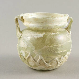
Oak root, light micrograph

![]()
Wall Art and Photo Gifts from Science Photo Library
Oak root, light micrograph
Oak root. Light micrograph of a section through a secondary root from an oak (Quercus sp.) tree. The primary cortex (outer layer) has been shed by the formation of a circular meristem, the periderm, which forms an outer layer of cork (phellum, black). Under the cork layer is a thin cortex (blue) and rings of several phloem layers (light blue) with patches of fibres (red). Next is the ring of cambium tissue (light blue), which contains an outer layer of phloem vessels and an inner layer of xylem (centre). The wood is classified as diffuse-porous, with large vessels and small cells of xylem parenchyma (light red). Six big rays (light blue) and small rays (blackish) of primary xylem tissue radiate from the centre. Magnification: x13 when printed 10
Science Photo Library features Science and Medical images including photos and illustrations
Media ID 6338725
© DR KEITH WHEELER/SCIENCE PHOTO LIBRARY
Cell Biology Cork Cortex Cytological Cytology Dicot Dicots Dicotyledon Dicotyledons Fibre Fibres Histological Histology Layer Layers Meristem Microscopy Parenchyma Phloem Primary Xylem Root Stain Stained Structural Structures Tissue Vessel Vessels Wood Xylem Cells Light Micrograph Light Microscope Section Sectioned
EDITORS COMMENTS
This print showcases the intricate beauty of an oak root, as seen through a light micrograph. The image provides a detailed glimpse into the inner workings of this secondary root from a majestic oak tree (Quercus sp. ). The outer layer, known as the primary cortex, has been shed to make way for the formation of a circular meristem called periderm. This periderm acts as an outer layer of cork, depicted here in striking black. Beneath the cork layer lies a thin cortex in mesmerizing shades of blue, surrounded by rings of phloem layers in lighter hues. These phloem layers are interspersed with patches of vibrant red fibers that add texture and depth to the composition. Moving inward, we encounter the ring-shaped cambium tissue also colored in light blue tones. Within this tissue reside both an outer layer composed of phloem vessels and an inner layer made up of xylem cells at its center. The wood structure visible here is classified as diffuse-porous due to its large vessels and small cells comprising xylem parenchyma showcased in delicate shades of light red. Radiating from the center are six prominent rays colored in soft blue along with smaller rays appearing blackish. This stunning photograph offers viewers a fascinating insight into the complex cellular architecture and botanical wonders found within nature's creations.
MADE IN THE USA
Safe Shipping with 30 Day Money Back Guarantee
FREE PERSONALISATION*
We are proud to offer a range of customisation features including Personalised Captions, Color Filters and Picture Zoom Tools
SECURE PAYMENTS
We happily accept a wide range of payment options so you can pay for the things you need in the way that is most convenient for you
* Options may vary by product and licensing agreement. Zoomed Pictures can be adjusted in the Cart.