Retina structure

![]()
Wall Art and Photo Gifts from Science Photo Library
Retina structure
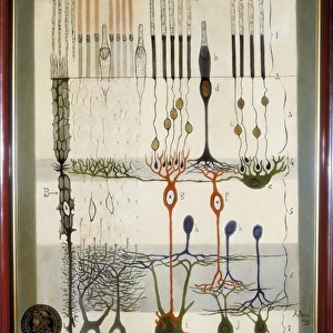
Retinal structure, artwork. Light falling on the retina passes from top to bottom. At bottom is the choroid layer (red), which lines the inside of the eye. At the back of the eye the choroid is covered by the retina. The inner layer of the retina, the retinal pigment epithelium (white) prevents light passing out of the eye. Light sensitive photoreceptor cells form the next layer and are of 2 types; rods (green) aid vision in dim light and cones (purple) allow colour vision. Light triggers electrical impulses in the interneurones (nerve cells, blue, orange and red). The impulses are passed onto the ganglion nerve cells (grey) and then the brain. At centre is a Muller cell (yellow), a support cell for the neurones
Science Photo Library features Science and Medical images including photos and illustrations
Media ID 6346445
© FRANCIS LEROY, BIOCOSMOS/SCIENCE PHOTO LIBRARY
Choroid Cones Ganglion Glial Cell Labeled Labelled Labels Nerve Fibres Nerves Neurone Neurones Neurons Retina Retinal Rods Sense Sight Text Vision Visual Nervous System
EDITORS COMMENTS
This print showcases the intricate structure of the retina, offering a mesmerizing glimpse into the inner workings of our visual system. Against a striking black background, this artwork highlights the complexity and beauty found within our eyes. At first glance, we are drawn to the vibrant colors that bring life to this illustration. The choroid layer, depicted in vivid red, lines the inside of the eye and acts as a protective shield for deeper structures. Just above it lies the retinal pigment epithelium, portrayed in pristine white, which plays a crucial role in preventing light from escaping out of our eyes. Moving further inward, we encounter an array of light-sensitive photoreceptor cells that form distinct layers within the retina. The rods, represented by soothing shades of green, aid us in seeing clearly even under dim lighting conditions. In contrast, cones take on an enchanting purple hue and enable us to perceive vibrant colors with remarkable precision. The interneurons elegantly weave their way through this intricate network like delicate threads of blue, orange and red. These nerve cells serve as messengers between different layers of neurons before transmitting electrical impulses to ganglion nerve cells – depicted here in calming grey tones – which ultimately relay visual information to our brain for processing. Amidst this symphony of neural activity stands a Muller cell painted in radiant yellow—a steadfast support system for these remarkable neurons that make sight possible. In sumptuous detail and meticulous labeling throughout its composition, this artful depiction invites us to marvel at nature's masterpiece—the human eye—and appreciate both its biological brilliance and aesthetic allure.
MADE IN THE USA
Safe Shipping with 30 Day Money Back Guarantee
FREE PERSONALISATION*
We are proud to offer a range of customisation features including Personalised Captions, Color Filters and Picture Zoom Tools
SECURE PAYMENTS
We happily accept a wide range of payment options so you can pay for the things you need in the way that is most convenient for you
* Options may vary by product and licensing agreement. Zoomed Pictures can be adjusted in the Cart.