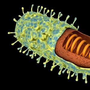
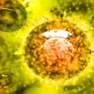
Semliki Forest virus, computer model

![]()
Wall Art and Photo Gifts from Science Photo Library
Semliki Forest virus, computer model
Semliki Forest virus (SFV), computer model. This image was created using UCSF Chimera molecular modelling software and data from cryo-electron microscopy. It shows the outer (translucent grey) layer sliced in half and coloured to reveal low to high electron densities (red to blue). The inner layer (blue, green, pink) is the virus protein shell, known as the capsid. SFV is transmitted by mosquitoes and can cause encephalitis in humans and animals. It is also used as a tool in gene therapy and drug research. Cryo-electron microscopy uses beams of electrons, which are fired at multiple angles, to image specimens kept at minus 150 degrees Celsius. The slices of data are reconstructed into 3-D models on computer
Science Photo Library features Science and Medical images including photos and illustrations
Media ID 6412776
© UCSF CHIMERA/SCIENCE PHOTO LIBRARY
3 D Electron Microscopy 3 D Visualisation 3 D Visualization Alphavirus Capsid Chimera Computer Graphic Computer Rendering Cryo Electron Cryo Electron Microscope Cryo Em Cryoelectron Microscopy Cut Away Electron Cryomicroscopy Electron Density Electron Microscopy Infectious Disease Macromolecular Macromolecule Modelling Molecular Imaging Particle Protein Database Reconstruction Shape Structural Biology Surface Ucsf Chimera Vector Viral Virion Virology Viruses Genetics Micro Biology Microbiological Molecular Model Molecular Structure Pathogen Protein Virus
EDITORS COMMENTS
This print showcases a computer model of the Semliki Forest virus (SFV), created using UCSF Chimera molecular modelling software and data obtained from cryo-electron microscopy. The image provides a unique glimpse into the intricate structure of this pathogenic virus. The translucent grey outer layer, sliced in half and color-coded to reveal electron densities ranging from red to blue, highlights the complexity of SFV's composition. Within this outer layer lies the virus protein shell, known as the capsid, depicted in shades of blue, green, and pink. SFV is primarily transmitted through mosquito bites and can lead to encephalitis in both humans and animals. However, beyond its role as a harmful pathogen, SFV also serves as a valuable tool in gene therapy research and drug development. Cryo-electron microscopy played an essential role in capturing this detailed representation. By bombarding specimens with beams of electrons at various angles while maintaining temperatures as low as minus 150 degrees Celsius, scientists were able to reconstruct these slices into three-dimensional models on a computer screen. This stunning visual not only sheds light on the structural biology of SFV but also emphasizes the significance of advanced imaging techniques like cryo-electron microscopy for understanding infectious diseases at their molecular level.
MADE IN THE USA
Safe Shipping with 30 Day Money Back Guarantee
FREE PERSONALISATION*
We are proud to offer a range of customisation features including Personalised Captions, Color Filters and Picture Zoom Tools
SECURE PAYMENTS
We happily accept a wide range of payment options so you can pay for the things you need in the way that is most convenient for you
* Options may vary by product and licensing agreement. Zoomed Pictures can be adjusted in the Cart.