Home > Europe > United Kingdom > Scotland > Moray > Keith
Spongy bone, light micrograph

![]()
Wall Art and Photo Gifts from Science Photo Library
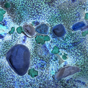
Spongy bone, light micrograph
Spongy bone. Light micrograph of a section through stained and decalcified human spongy bone. This bone type is also called cancellous bone. The bone matrix is composed of collagen fibres with lacunae containing osteocytes, which maintain the bone structure. Spongy bone has trabeculae which act as a series of girders, which transfer the forces received at the end of the bone, to the stronger outer walls of compact bone. The cavities in the spongy bone and central bone marrow are the site of blood cell formation and full of blood vessels. Magnification: x36 when printed at 10 centimetres across
Science Photo Library features Science and Medical images including photos and illustrations
Media ID 6279049
© DR KEITH WHEELER/SCIENCE PHOTO LIBRARY
Bone Marrow Cancellous Bone Circle Circular Collagen Fibre Cross Section Fibres Histological Histology Lacuna Lacunae Osteocyte Osteocytes Osteology Round Spongy Bone Structural Trabecula Transverse Artery Light Micrograph Light Microscope Section Sectioned
EDITORS COMMENTS
This print showcases the intricate structure of spongy bone, also known as cancellous bone, in a stained and decalcified human sample. The light micrograph reveals a section through this remarkable bone type, highlighting its unique characteristics. Composed of collagen fibers with lacunae housing osteocytes, which play a crucial role in maintaining the bone's structure, spongy bone exhibits an awe-inspiring circular pattern. Trabeculae within the spongy bone act like sturdy girders that efficiently transfer forces from the end of the bone to the stronger outer walls made up of compact bone. These cavities are not only responsible for blood cell formation but are also filled with an intricate network of blood vessels. At a magnification of x36 when printed at 10 centimeters across, this image provides us with a glimpse into the fascinating world of biology and anatomy. It serves as a reminder of our own structural complexity and highlights how essential healthy bones are for our overall well-being. The detailed histological features captured by this light microscope image offer valuable insights into osteology and provide scientists with important information about various aspects related to skeletal health. This visually stunning photograph is sure to captivate both scientific enthusiasts and those fascinated by the wonders hidden within our bodies.
MADE IN THE USA
Safe Shipping with 30 Day Money Back Guarantee
FREE PERSONALISATION*
We are proud to offer a range of customisation features including Personalised Captions, Color Filters and Picture Zoom Tools
SECURE PAYMENTS
We happily accept a wide range of payment options so you can pay for the things you need in the way that is most convenient for you
* Options may vary by product and licensing agreement. Zoomed Pictures can be adjusted in the Cart.