Home > Europe > United Kingdom > Scotland > Moray > Keith
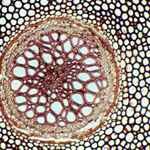
Water fern rhizome, light micrograph

![]()
Wall Art and Photo Gifts from Science Photo Library
Water fern rhizome, light micrograph
Water fern rhizome. Polarised light micrograph of a section through a rhizome from a water fern (Marselia vestita). Under the epidermis (the outer layer) is the cortex with lamellae plates enclosing large air spaces (lacunae) which store oxygen and are characteristic of hydrophytes. The inner cortex includes a ring of thick sclerotic supporting cells (red). Inside this is a central vascular cylinder that includes endoderm, phloem (light pink), pericycle (deep blue) and xylem (yellow-pink) tissues, around a central pith (red). Magnification: x44 when printed at 10 centimetres across
Science Photo Library features Science and Medical images including photos and illustrations
Media ID 6334875
© DR KEITH WHEELER/SCIENCE PHOTO LIBRARY
Air Spaces Cellular Circle Circular Cortex Cross Section Endoderm Fern Hydrophyte Hydrophytic Lacuna Lacunae Nutrient Transport Pericycle Phloem Pith Polarised Light Micrograph Polarised Light Microscopy Polarized Rhizome Round Tissue Transverse Vascular Bundle Vascular Bundles Water Transport Xylem Cells Hydrophytes Light Micrograph Light Microscope Section Sectioned
FEATURES IN THESE COLLECTIONS
> Animals
> Mammals
> Muridae
> Water Mouse
> Arts
> Artists
> L
> polarized light
> Arts
> Realistic drawings
> Nature art
> Botanical artwork
> Europe
> United Kingdom
> Scotland
> Moray
> Keith
EDITORS COMMENTS
This print showcases the intricate beauty of a water fern rhizome, captured through a polarised light micrograph. The image reveals the complex structure of this hydrophytic plant's underground stem, known as the rhizome. The outer layer, or epidermis, is visible in this circular section. Just beneath it lies the cortex, which contains lamellae plates enclosing large air spaces called lacunae. These unique structures serve as oxygen stores and are characteristic of plants that thrive in aquatic environments. Moving inward, we encounter a ring of thick sclerotic supporting cells colored in vibrant red. This inner cortex surrounds a central vascular cylinder composed of various tissues including endoderm, phloem (light pink), pericycle (deep blue), and xylem (yellow-pink). These components play crucial roles in nutrient transport and water circulation within the plant. At the very center lies the pith, also depicted in red. Together with all these elements, they form an intricate network responsible for sustaining growth and survival. With its magnification at 44 times when printed at 10 centimeters across, this stunning photograph allows us to appreciate nature's complexity on a microscopic level. It serves as a reminder of how even seemingly small organisms can possess remarkable intricacy and beauty worth exploring further through scientific inquiry.
MADE IN THE USA
Safe Shipping with 30 Day Money Back Guarantee
FREE PERSONALISATION*
We are proud to offer a range of customisation features including Personalised Captions, Color Filters and Picture Zoom Tools
SECURE PAYMENTS
We happily accept a wide range of payment options so you can pay for the things you need in the way that is most convenient for you
* Options may vary by product and licensing agreement. Zoomed Pictures can be adjusted in the Cart.