Fine Art Print : Foot anatomy, artwork


![]()
Fine Art Prints from Science Photo Library
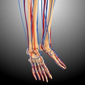
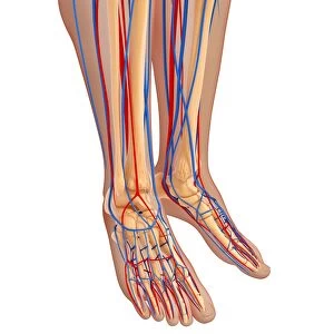
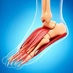
Foot anatomy, artwork
Foot anatomy, computer artwork
Science Photo Library features Science and Medical images including photos and illustrations
Media ID 9258079
© PIXOLOGICSTUDIO/SCIENCE PHOTO LIBRARY
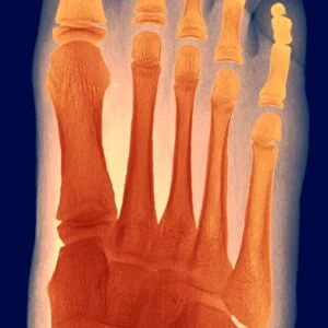
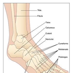
Foot Lymphatic System Metatarsal Phalanx Tarsal Vascular System Artery Blood Supply Blood Vessel Blue Background Cardiovascular System Circulatory System Nervous System Vein
20"x16" (+3" Border) Fine Art Print
Discover the intricacies of the human foot with our Fine Art Print from Media Storehouse, featuring this stunning computer-generated artwork by Science Photo Library. This captivating foot anatomy image showcases the complex network of bones, muscles, and tendons that make up this essential part of the human body. Bring the wonders of science into your home or office with this beautiful and educational print. Perfect for healthcare professionals, students, or anyone with an appreciation for the marvels of the human body.
20x16 image printed on 26x22 Fine Art Rag Paper with 3" (76mm) white border. Our Fine Art Prints are printed on 300gsm 100% acid free, PH neutral paper with archival properties. This printing method is used by museums and art collections to exhibit photographs and art reproductions.
Our fine art prints are high-quality prints made using a paper called Photo Rag. This 100% cotton rag fibre paper is known for its exceptional image sharpness, rich colors, and high level of detail, making it a popular choice for professional photographers and artists. Photo rag paper is our clear recommendation for a fine art paper print. If you can afford to spend more on a higher quality paper, then Photo Rag is our clear recommendation for a fine art paper print.
Estimated Image Size (if not cropped) is 40.6cm x 50.8cm (16" x 20")
Estimated Product Size is 55.9cm x 66cm (22" x 26")
These are individually made so all sizes are approximate
Artwork printed orientated as per the preview above, with portrait (vertical) orientation to match the source image.
EDITORS COMMENTS
This print showcases the intricate and fascinating world of foot anatomy, beautifully depicted through computer artwork. The close-up view allows us to appreciate the detailed illustration of a human foot's skeletal structure, emphasizing its various components such as the toes, veins, and bones. Against a serene blue background, this artwork provides an educational insight into the healthy functioning of our feet. It serves as a reminder of how crucial understanding foot anatomy is for medicine and healthcare professionals in diagnosing and treating conditions related to this vital part of our body. The image highlights not only the bone structure but also includes important elements like blood vessels and nerves that contribute to the overall functionality of our feet. By showcasing tarsal bones, metatarsals, phalanges, arteries, and other components associated with both circulatory systems (cardiovascular system and lymphatic system), it offers a comprehensive representation of foot anatomy. With its attention to detail and accuracy in depicting human representation within medical illustrations, this artwork from Science Photo Library exemplifies their commitment to providing valuable resources for scientific education. Whether you are studying or simply intrigued by human anatomy or healthcare practices related to leg anatomy specifically - this visually striking print will surely captivate your interest.
MADE IN THE USA
Safe Shipping with 30 Day Money Back Guarantee
FREE PERSONALISATION*
We are proud to offer a range of customisation features including Personalised Captions, Color Filters and Picture Zoom Tools
SECURE PAYMENTS
We happily accept a wide range of payment options so you can pay for the things you need in the way that is most convenient for you
* Options may vary by product and licensing agreement. Zoomed Pictures can be adjusted in the Cart.