Fine Art Print > Science > SEM
Fine Art Print : Nerve cells, SEM


![]()
Fine Art Prints from Science Photo Library
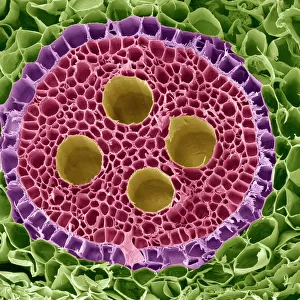
Nerve cells, SEM
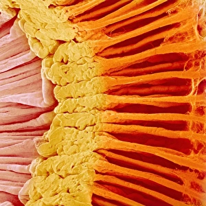
Nerve cells. Coloured scanning electron micrograph (SEM) of nerve cells, known as neurones. Nerve cells occur in the brain, spinal cord, and in ganglia. Each nerve cell has a large cell body (brown) with several long processes extending from it. The processes usually consist of one thicker axon and several thinner branched dendrites. The dendrites collect information in the form of nerve impulses from other nerve cells and pass it to the cell body. This information is interpreted by the cell body, and then passed on along the axon. In this way neurones allow information to be rapidly received, interpreted, and relayed around the body. Magnification: x800 when printed at 10 centimetres wide
Science Photo Library features Science and Medical images including photos and illustrations
Media ID 10826114
© STEVE GSCHMEISSNER/SCIENCE PHOTO LIBRARY
Axon Cell Body Dendrite Dendrites Magnified Image Microscopic Photos Nerve Cell Nervous Neurone Neurones Subjects System Team Teams Teamwork Togetherness Cells Images Together
20"x16" (+3" Border) Fine Art Print
Discover the intricacy of life with our Fine Art Prints from Media Storehouse. This captivating image showcases the beauty of Nerve Cells, as captured in stunning detail through Coloured Scanning Electron Micrograph (SEM) technology from Science Photo Library. Each print brings the complexity of the natural world into your home or office, serving as a conversation starter and a source of inspiration. Our high-quality prints are produced using archival inks and materials, ensuring your investment is a long-lasting one. Elevate your space with the mesmerizing visuals of our Fine Art Prints collection.
20x16 image printed on 26x22 Fine Art Rag Paper with 3" (76mm) white border. Our Fine Art Prints are printed on 300gsm 100% acid free, PH neutral paper with archival properties. This printing method is used by museums and art collections to exhibit photographs and art reproductions.
Our fine art prints are high-quality prints made using a paper called Photo Rag. This 100% cotton rag fibre paper is known for its exceptional image sharpness, rich colors, and high level of detail, making it a popular choice for professional photographers and artists. Photo rag paper is our clear recommendation for a fine art paper print. If you can afford to spend more on a higher quality paper, then Photo Rag is our clear recommendation for a fine art paper print.
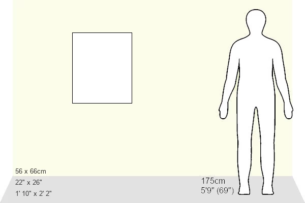
Estimated Image Size (if not cropped) is 40.6cm x 50.8cm (16" x 20")
Estimated Product Size is 55.9cm x 66cm (22" x 26")
These are individually made so all sizes are approximate
Artwork printed orientated as per the preview above, with portrait (vertical) orientation to match the source image.
FEATURES IN THESE COLLECTIONS
EDITORS COMMENTS
This print showcases the intricate beauty of nerve cells, also known as neurones. Coloured with vibrant hues, this scanning electron micrograph (SEM) captures the essence of these vital components found in the brain, spinal cord, and ganglia. Each nerve cell is characterized by a prominent cell body in rich brown tones, accompanied by numerous elongated processes extending from it. These processes consist of one thicker axon and several thinner branched dendrites that play crucial roles in information transmission within our nervous system. The dendrites act as collectors, receiving nerve impulses from other neighbouring nerve cells and relaying them to the cell body for interpretation. Once interpreted, this valuable information is swiftly passed along the axon. Neurones are fundamental to our ability to rapidly receive, interpret, and relay information throughout our bodies. They form an intricate network that enables seamless communication between different parts of our anatomy. This microscopic image magnified at x800 reveals their remarkable structure and highlights their significance in facilitating efficient neural communication. The mesmerizing complexity displayed in this photograph serves as a reminder of the incredible teamwork exhibited by these neurones within us. Together they function harmoniously to ensure smooth operation of our nervous system—a testament to the power of collaboration even at a cellular level.
MADE IN THE USA
Safe Shipping with 30 Day Money Back Guarantee
FREE PERSONALISATION*
We are proud to offer a range of customisation features including Personalised Captions, Color Filters and Picture Zoom Tools
SECURE PAYMENTS
We happily accept a wide range of payment options so you can pay for the things you need in the way that is most convenient for you
* Options may vary by product and licensing agreement. Zoomed Pictures can be adjusted in the Cart.