Framed Print : Enterocyte, TEM


![]()
Framed Photos from Science Photo Library
Enterocyte, TEM
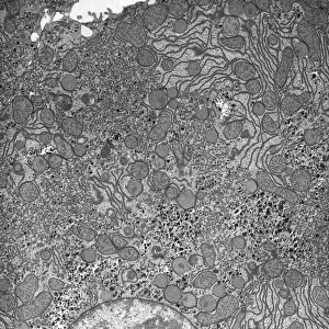
Enterocyte. Transmission electron micrograph (TEM) of a section through the cytoplasm and part of the nucleus of an enterocyte cell located in a crypt of Lieberkuhn of the small intestine, showing an abundance of rough endoplasmic reticulum (dark lines), Golgi membranes ((semi-circular lines) and vesicles (dark circles). Crypt enterocytes secrete intestinal fluid, an electrolyte solution that is taken up by the enterocytes of the villous epithelium and assists with their absorption of nutrients from the gut. Crypts of Lieberkuhn contain the intestinal epithelial stem cells that constantly proliferate to renew the enterocytes lost from the upper regions of the villi. Magnification: x20, 000 when printed 10 centimetres tall
Science Photo Library features Science and Medical images including photos and illustrations
Media ID 9241543
© MICROSCAPE/SCIENCE PHOTO LIBRARY
Black And White Bowel Bowels Cell Biology Cytological Cytology Cytoplasm Digestive System Enterocyte Enterocytes Gastrointestinal Tract Histological Histology Intestines Membranes Nucleus Organelle Organelles Secretory Small Intestine Transmission Electron Micrograph Transmission Electron Microscope Vesicle Vesicles Cells Section Sectioned
12"x10" Modern Frame
Discover the intricacies of the microscopic world with Media Storehouse's Framed Prints. Feast your eyes on this stunning Transmission Electron Micrograph (TEM) of an enterocyte cell, captured by Science Photo Library. Witness the intricate details of this intestinal cell's cytoplasm and nucleus, as seen through the lens of a transmission electron microscope. Our Framed Prints are not just decorative pieces, they're windows into the wonders of science. Bring this captivating image into your home or office and ignite curiosity and conversation. Order yours today!
10x8 Print in an MDF Wooden Frame with 180 gsm Satin Finish Paper. Glazed using shatter proof thin plexi glass. Frame thickness is 1 inch and depth 0.75 inch. Fluted cardboard backing held with clips. Supplied ready to hang with sawtooth hanger and rubber bumpers. Spot clean with a damp cloth. Packaged foam wrapped in a card.
Contemporary Framed and Mounted Prints - Professionally Made and Ready to Hang
Estimated Image Size (if not cropped) is 25.4cm x 25.4cm (10" x 10")
Estimated Product Size is 25.4cm x 30.5cm (10" x 12")
These are individually made so all sizes are approximate
Artwork printed orientated as per the preview above, with landscape (horizontal) or portrait (vertical) orientation to match the source image.
EDITORS COMMENTS
This print showcases the intricate details of an enterocyte cell, as seen through a transmission electron microscope (TEM). The image reveals the complex structure of the cytoplasm and nucleus, highlighting various organelles and membranes. One prominent feature is the abundance of rough endoplasmic reticulum, represented by dark lines throughout the cell. Additionally, semi-circular Golgi membranes can be observed alongside numerous vesicles in the form of dark circles. Enterocytes located in crypts of Lieberkuhn within the small intestine play a crucial role in secreting intestinal fluid, which aids in electrolyte balance. This fluid is then absorbed by enterocytes present on villous epithelium to facilitate nutrient absorption from digested food. Moreover, these crypts house intestinal epithelial stem cells that constantly divide to replenish lost enterocytes from upper regions of villi. With a magnification level of x20,000 when printed at 10 centimeters tall, this photograph offers a remarkable glimpse into cellular biology and histology. It provides valuable insights into how our digestive system functions at a microscopic level. Science enthusiasts and researchers alike will appreciate this monochrome masterpiece captured by Science Photo Library's advanced TEM technology.
MADE IN THE USA
Safe Shipping with 30 Day Money Back Guarantee
FREE PERSONALISATION*
We are proud to offer a range of customisation features including Personalised Captions, Color Filters and Picture Zoom Tools
SECURE PAYMENTS
We happily accept a wide range of payment options so you can pay for the things you need in the way that is most convenient for you
* Options may vary by product and licensing agreement. Zoomed Pictures can be adjusted in the Cart.