Framed Print > Arts > Artists > Leonardo da Vinci > Anatomy studies by Leonardo da Vinci
Framed Print : Skull anatomy by Leonardo da Vinci


Framed Photos from Science Photo Library
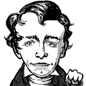
Skull anatomy by Leonardo da Vinci
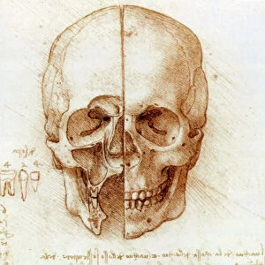
Skull anatomy by Leonardo da Vinci. Historical artwork and notes on the anatomy of the human skull and teeth, by the Italian artist and scientist Leonardo da Vinci (1452-1519). This bisected skull shows the external structure (right), and dissected facial sinuses (left), the air-filled spaces inside the bones of the face. The diagram at lower left shows the teeth present in one half of the mouth: 4 incisors, 2 canines, 4 pre-molars, and 6 molars. Da Vinci was the first anatomist known to have correctly noted the number and root structure of human teeth. The notes are an example of his mirror writing, which was written backwards from right to left, and could be read in a mirror
Science Photo Library features Science and Medical images including photos and illustrations
Media ID 6419516
© SHEILA TERRY/SCIENCE PHOTO LIBRARY
1400s 1500s 15th Century 16th Century Accurate Black And White Bones Canine Canines Cavities Cavity Code Cranium Dental Diagram Dissected Dissection Drawing Face Facial Front Frontal Incisor Incisors Italian Jaws Leonardo Da Vinci Mandible Maxilla Maxillary Molar Molars Monochrome Image Mouth Nasal Nose Note Note Book Oral Orbits Paper Pencil People Person Persons Renaissance Root Roots Sinus Sinuses Skeletal Socket Sockets Teeth Tooth Writing Zygomatic Arch Annotation Backwards Mirror Writing Sphenoid
12"x10" Modern Frame
Introducing the Media Storehouse Framed Prints featuring the iconic "Skull anatomy by Leonardo da Vinci" from Science Photo Library. This exquisite artwork showcases the masterful mind of Leonardo da Vinci, an Italian artist and scientist, as he meticulously documented the intricacies of human skull and teeth anatomy. Each print is expertly framed to preserve and enhance the beauty of this historical masterpiece. Bring the brilliance of Leonardo's scientific exploration into your home or office, and elevate your space with this timeless piece of art and history.
10x8 Print in an MDF Wooden Frame with 180 gsm Satin Finish Paper. Glazed using shatter proof thin plexi glass. Frame thickness is 1 inch and depth 0.75 inch. Fluted cardboard backing held with clips. Supplied ready to hang with sawtooth hanger and rubber bumpers. Spot clean with a damp cloth. Packaged foam wrapped in a card.
Contemporary Framed and Mounted Prints - Professionally Made and Ready to Hang
Estimated Image Size (if not cropped) is 25.4cm x 25.4cm (10" x 10")
Estimated Product Size is 30.5cm x 25.4cm (12" x 10")
These are individually made so all sizes are approximate
Artwork printed orientated as per the preview above, with landscape (horizontal) or portrait (vertical) orientation to match the source image.
FEATURES IN THESE COLLECTIONS
> Arts
> Art Movements
> Renaissance Art
> Arts
> Artists
> Leonardo da Vinci
> Anatomy studies by Leonardo da Vinci
> Arts
> Artists
> Leonardo da Vinci
> Renaissance art
> Arts
> Artists
> Leonardo da Vinci
> Sketches and drawings by Leonardo da
> Arts
> Artists
> Leonardo da Vinci
> Arts
> Artists
> Leonardo Da Vinci
> Arts
> Artists
> V
> Leonardo da Vinci
> Arts
> Minimalist artwork
> Monochrome artwork
> Black and white artwork
> Arts
> Minimalist artwork
> Monochrome artwork
> Fine art
> Arts
> Minimalist artwork
> Monochrome artwork
> Monochrome paintings
> Popular Themes
> Leonardo da Vinci
> Posters
> Scientific Posters
> Science Photo Library
> History
EDITORS COMMENTS
This print showcases the remarkable "Skull anatomy" by Leonardo da Vinci, a true masterpiece of historical artwork and scientific exploration. Created in the 16th century, this illustration provides an intricate view of the human skull and teeth, meticulously dissected by the brilliant Italian artist and scientist. Leonardo's attention to detail is evident as he presents both the external structure of the skull on the right side and reveals the inner facial sinuses on the left. These air-filled spaces within our facial bones are beautifully depicted through his skillful pencil strokes. One notable aspect of this artwork is Leonardo's groundbreaking understanding of dental anatomy. In a diagram at lower left, he accurately notes each type of tooth present in one half of a mouth: incisors, canines, pre-molars, and molars. This revelation marked him as one of history's first anatomists to correctly identify not only their number but also their root structure. The accompanying annotations are written in Leonardo's signature mirror writing style – backwards from right to left – which could be deciphered using a mirror. This unique code adds an intriguing element to his work while showcasing his exceptional intellect. As we admire this rare glimpse into Leonardo da Vinci's genius mind, we are reminded once again why he remains an icon even centuries after his time. His dedication to accuracy and innovation continues to inspire scientists, artists, and thinkers alike across generations.
MADE IN THE USA
Safe Shipping with 30 Day Money Back Guarantee
FREE PERSONALISATION*
We are proud to offer a range of customisation features including Personalised Captions, Color Filters and Picture Zoom Tools
SECURE PAYMENTS
We happily accept a wide range of payment options so you can pay for the things you need in the way that is most convenient for you
* Options may vary by product and licensing agreement. Zoomed Pictures can be adjusted in the Cart.