Abnormal Collection (#55)

"Unveiling the Abnormal: A Glimpse into Extraordinary Phenomena" Gas pillars in the Eagle Nebula: Witness the mesmerizing dance of celestial gas pillars

Background imageAbnormal Collection: Wound healing, artwork

Wound healing, artwork
Wound healing. Artwork 2 of 4 showing wound healing. This is the proliferative phase, which occurs between 2 to 5 days after the formation of the wound

Background imageAbnormal Collection: Childhood rickets, X-ray

Childhood rickets, X-ray
Childhood rickets. Coloured frontal X-ray of the legs of a 3-year-old child with rickets. Rickets (Osteomalacia) occurs predominantly in children

Background imageAbnormal Collection: Broken hip, X-ray

Broken hip, X-ray

Background imageAbnormal Collection: Arteritis of the knee, X-ray

Arteritis of the knee, X-ray
Arteritis of the knee. Coloured X-ray image of the knee of an 85-year-old patient with arteritis. Arteritis is an inflammation of the walls of arteries

Background imageAbnormal Collection: Calcified aortic artery, 3D CT scan

Calcified aortic artery, 3D CT scan
Coloured three-dimensional (3D) Computed Tomography (CT) scan in front view of the abdomen in a patient showing the kidneys (red)

Background imageAbnormal Collection: Tooth decay, X-ray

Tooth decay, X-ray
Tooth decay. Coloured X-ray of a profile view of a 22-year-old patients teeth, showing a cavity (decay) in the left lower molar (light grey, centre left)

Background imageAbnormal Collection: Small bowel obstruction, X-ray

Small bowel obstruction, X-ray

Background imageAbnormal Collection: Abdominal air pockets, X-ray

Abdominal air pockets, X-ray

Background imageAbnormal Collection: Emphysema, 3D CT scan

Emphysema, 3D CT scan

Background imageAbnormal Collection: Takayasus arteritis, 3d CT scan

Takayasus arteritis, 3d CT scan
Takayasus arteritis. Coloured 3d computed tomography (CT) angiogram of the arteries of the neck and head of a 56-year-old patient with Takayasus arteritis

Background imageAbnormal Collection: Tourettes treatment, X-ray

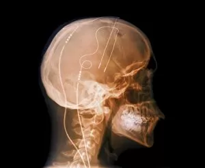
Tourettes treatment, X-ray
Tourettes treatment, coloured X-ray. Profile view of the skull of a 25-year-old man with Tourettes syndrome, showing electrodes implanted in his brain
Background imageAbnormal Collection: Damaged knee, artwork

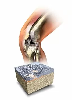
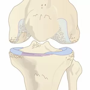
Damaged knee, artwork
Damaged knee. Artwork showing the internal anatomy of a damaged human knee, with an inset (bottom) of the cartilage (grey) that covers the ends of the bones forming this joint

Background imageAbnormal Collection: Aortic aneurysm, X-ray

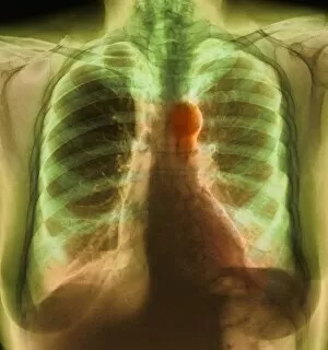
Aortic aneurysm, X-ray
Aortic aneurysm. Coloured X-ray of the chest of a 68-year-old woman with an aortic aneurysm. An aneurysm is a swelling of an artery where the wall has weakened, here on the arch where the aorta

Background imageAbnormal Collection: Stenosis of carotid artery, CT scan

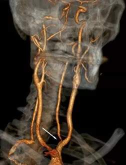
Stenosis of carotid artery, CT scan
Stenosis of carotid artery. Coloured three-dimensional computed tomography (CT) angiography scan of the neck arteries (red) of a 41-year-old patient, showing a stenosis (narrowing)
Background imageAbnormal Collection: Osteoporosis, artwork

Osteoporosis, artwork
Osteoporosis. Artwork of vertical sections comparing the upper parts of a healthy femur (left) and one with reduced bone mass (right)
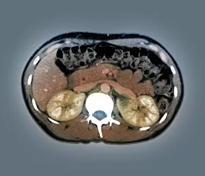
Background imageAbnormal Collection: Medullary sponge kidney, CT scan

Medullary sponge kidney, CT scan
Medullary sponge kidney. Coloured computed tomography (CT) scan in axial section of the abdomen of a 25-year-old patient with medullary sponge kidney, or Cacchi Ricci disease

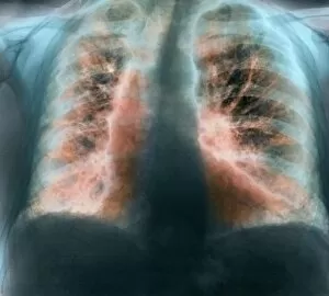
Background imageAbnormal Collection: Fibrosing alveolitis, X-ray

Fibrosing alveolitis, X-ray
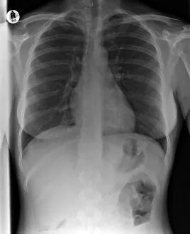
Fibrosing alveolitis. Coloured X-ray of the chest of an 82-year-old man with fibrosing alveolitis. Also known as idiopathic pulmonary fibrosis and cryptogenic fibrosing alveolitis

Background imageAbnormal Collection: Shrapnel injury, X-ray

Shrapnel injury, X-ray

Background imageAbnormal Collection: Healthy and crenated red blood cells, SEM

Healthy and crenated red blood cells, SEM
Red blood cells. Coloured scanning electron micrograph (SEM) of normal healthy red blood cells (red) and a crenated red blood cell (white)

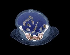
Background imageAbnormal Collection: Chronic liver disease, CT scan

Chronic liver disease, CT scan
Chronic liver disease. Coloured computed tomography (CT) scan of an axial section through the abdominopelvic region of a 58-year-old patient with a history of hepatitis C

Background imageAbnormal Collection: Slipped disc, MRI scan

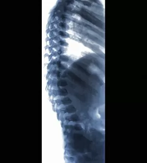
Slipped disc, MRI scan
Slipped disc, coloured magnetic resonance imaging (MRI) scan. Profile view of the spine of a 59-year-old patient with severe rheumatoid arthritis and a herniated (slipped) disc (red)

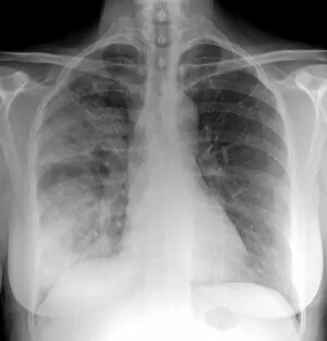
Background imageAbnormal Collection: Pneumonia, X-ray

Pneumonia, X-ray

Background imageAbnormal Collection: Broken jawbone, X-ray

Broken jawbone, X-ray
Broken jawbone. X-ray of the jaw of a young female patient with a fractured mandible (lower jawbone). Two cracks can be seen in the bone, at right and bottom left

Background imageAbnormal Collection: Arteritis, 3d CT scan

Arteritis, 3d CT scan
Arteritis. Coloured 3d computed tomography (CT) angiogram of the abdominal aorta in a 60-year-old patient after treatment for arteritis

Background imageAbnormal Collection: Aortic aneurysm CT scan

Aortic aneurysm CT scan
Aortic aneurysm, coloured 3D computed tomography (CT) scan. Side view of the abdomen of a 65-year-old patient with an extended aneurysm of the aorta

Background imageAbnormal Collection: Charcot arthropathy, MRI scans

Charcot arthropathy, MRI scans
Charcot arthropathy. Coloured magnetic resonance imaging (MRI) scans of the foot of a 33-year-old patient with Charcot arthropathy

Background imageAbnormal Collection: Bone infection, X-ray

Bone infection, X-ray

Background imageAbnormal Collection: Spine in Hurler Syndrome, X-ray

Spine in Hurler Syndrome, X-ray
Spine in Hurler Syndrome. Coloured X-ray of the spine of a 26-month-old child with Hurler syndrome (also called mucopolysaccharidosis type I, MPS I)

Background imageAbnormal Collection: Swallowed toothbrush, X-ray

Swallowed toothbrush, X-ray
Swallowed toothbrush. X-ray of the abdomen of a female patient who has swallowed a toothbrush (lower middle)

Background imageAbnormal Collection: Osteoarthritis of the knee, artwork

Osteoarthritis of the knee, artwork
Osteoarthritis of the knee. Computer artwork showing a knee joint affected by osteoarthritis. Osteoarthritis is a degenerative disease that involves degradation of the bone

Background imageAbnormal Collection: Abdominal aortic aneurysm, artwork

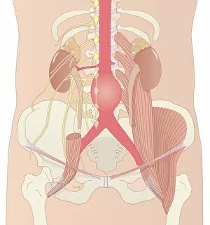
Abdominal aortic aneurysm, artwork
Abdominal aortic aneurysm. Computer artwork showing a large aneurysm (round, centre) in the abdominal section of the aorta

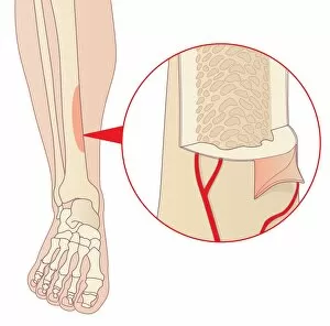
Background imageAbnormal Collection: Shin splints, artwork

Shin splints, artwork
Shin splints. Computer artwork of a lower leg showing an area of the tibia (shin bone) affected by medial tibial stress syndrome, or shin splints

Background imageAbnormal Collection: Dehydration, artwork

Dehydration, artwork
Dehydration. Computer artwork of a senior patient in a hospital bed, showing the signs of dehydration. These include: dry eyes and mouth; drowsiness

Background imageAbnormal Collection: Slipped disc, artwork

Slipped disc, artwork
Slipped disc. Artwork showing a slipped disc (orange) in a womans back. This condition is where one of the intervertebral discs (grey) in the backbone ruptures

Background imageAbnormal Collection: Normal and slipped disc, artwork

Normal and slipped disc, artwork
Normal and slipped disc. Artwork showing a normal (top) and slipped (bottom) disc in the backbone, seen from above. The rear of the backbone (with protruding processes)

Background imageAbnormal Collection: Parkinsons disease, artwork

Parkinsons disease, artwork
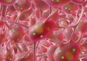
Parkinsons disease. Computer artwork of neurons (nerve cells, pink) containing Lewy bodies (green). Lewy bodies, which are deposits of protein

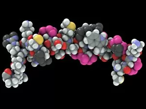
Background imageAbnormal Collection: Bovine prion protein, molecular model

Bovine prion protein, molecular model
Bovine prion protein. Computer model showing the molecular structure of a bovine (cow) prion protein. Prions are abnormal proteins that cause a group of fatal neurodegenerative diseases including BSE

Background imageAbnormal Collection: Capillary, artwork

Capillary, artwork
Capillary. Artwork showing the anatomical structure of a capillary seen in cross-section

Background imageAbnormal Collection: Abnormal sperm, SEM

Abnormal sperm, SEM
Abnormal sperm. Coloured scanning electron micrograph (SEM) of a sperm cell with an exposed central flagellum at the base of its tail

Background imageAbnormal Collection: Fractured pelvis, X-ray

Fractured pelvis, X-ray
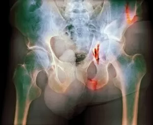
Fractured pelvis. Coloured X-ray of fractures (red) in the pelvis. The head of the femur (thigh bone) articulates with the acetabulum to form the hip joint

Background imageAbnormal Collection: Ectopic pancreatic stent, CT scan

Ectopic pancreatic stent, CT scan
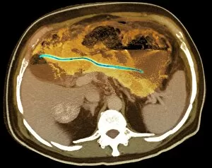
Ectopic pancreatic stent. Coloured computed tomography (CT) scan of an axial section through the abdomen of a patient, showing an ectopic (abnormally positioned) pancreatic stent (green)

Background imageAbnormal Collection: Fractured arm, X-ray

Fractured arm, X-ray
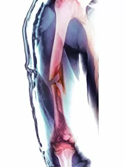
Fractured arm. Coloured X-ray of the front view of a fractured upper arm bone (humerus). The fracture is at centre (red), half way down the bone

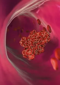
Background imageAbnormal Collection: Detached blood clot, artwork

Detached blood clot, artwork
Detached blood clot. Computer artwork of an embolus (detached blood clot) in a blood vessel. The erythrocytes (red blood cells) in the clot are held together by the protein fibrin (yellow lines)

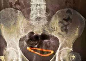
Background imageAbnormal Collection: Ectopic hydrocephalic shunt, X-ray

Ectopic hydrocephalic shunt, X-ray
Ectopic hydrocephalic shunt. Coloured X-ray of the pelvis of a patient, showing an ectopic (abnormally positioned) hydrocephalic shunt (orange)


For sale as Licensed Images

Choose your image, Select your licence and Download the media

"Unveiling the Abnormal: A Glimpse into Extraordinary Phenomena" Gas pillars in the Eagle Nebula: Witness the mesmerizing dance of celestial gas pillars, defying gravity and painting a surreal picture in the depths of space. Apollo 11 astronaut footprint on Moon: Behold an iconic abnormality, a single footprint etched by humanity's giant leap onto another world, forever marking our presence among the stars. Broken wrist bone, X-ray C017 / 7187: Delve into the intricate realm of human anatomy as we explore an abnormality captured within an X-ray image – a fractured wrist bone telling tales of resilience and healing. Solar prominence: Marvel at nature's fiery spectacle as solar prominences erupt from our nearest star, showcasing their abnormal beauty against the backdrop of infinite cosmic expanse. DAF 95 XF wide: Discover automotive innovation with this extraordinary truck model boasting exceptional width – its bold design challenges conventional norms to redefine road transport. HeLa cells, light micrograph C017 / 8299: Peer through a microscope lens to witness HeLa cells – immortalized yet controversial entities that have revolutionized medical research while raising ethical questions about their origin. Image of Jupiter taken with Hubble Telescope: Explore Jupiter's enigmatic atmosphere through an extraordinary image captured by Hubble Telescope; revealing swirling storms and vibrant bands that defy expectations. Knee pain, conceptual artwork: Dive into thought-provoking artistry depicting knee pain – a visual representation inviting contemplation on physical discomforts experienced by many but often hidden beneath societal masks. Joint pain, conceptual artwork: Engage your senses with evocative imagery portraying joint pain; transcending boundaries between art and reality to shed light on invisible struggles faced by individuals worldwide. Total hip replacement, X-ray.