Abnormal Collection (#53)

"Unveiling the Abnormal: A Glimpse into Extraordinary Phenomena" Gas pillars in the Eagle Nebula: Witness the mesmerizing dance of celestial gas pillars

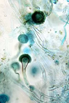
Background imageAbnormal Collection: Brown mould fungus, light micrograph

Brown mould fungus, light micrograph
Brown mould fungus. Light micrograph of a section through decaying plant tissue infected with brown mould fungus (Aspergillus glaucum)

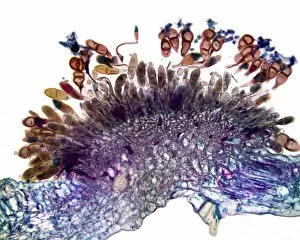
Background imageAbnormal Collection: Downy mildew infection, light micrograph

Downy mildew infection, light micrograph
Downy mildew infection. Light micrograph of a section through a grapevine (Vitis sp.) leaf infected with downy mildew (Plasmopara viticola)

Background imageAbnormal Collection: Anaemia, conceptual image

Anaemia, conceptual image. Computer artwork of red blood cells (erythrocytes) flowing through blood vessels (tubes). The red blood cells in the middle vessel have lost their colour

Background imageAbnormal Collection: Urethral stricture, artwork

Urethral stricture, artwork
Urethral stricture. Computer artwork of a narrowing of the urethra. This can be caused by injury or disease, such as urinary tract infections or other causes of urethritis

Background imageAbnormal Collection: Hollyhock rust fungus in a leaf

Hollyhock rust fungus in a leaf. Light micrograph of a section through a hollyhock (Alcea sp.) leaf infected with the parasitic Puccinia malvacearum rust, showing the teliospores (spores)

Background imageAbnormal Collection: Clubroot infection, light micrograph

Clubroot infection, light micrograph
Clubroot infection. Light micrograph a section through a cabbage root infected with clubroot (Plasmodiophora brassicae). Plasmodiophora is a parasitic protist that infects the roots of cabbages

Background imageAbnormal Collection: Pelvis in Pagets disease, X-ray

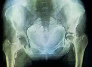
Pelvis in Pagets disease, X-ray
Coloured X-ray (front view) of the pelvis in a 60 year old patient showing the mottled and wispy appearance of the bones due to their increased porosity in Pagets disease

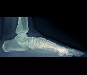
Background imageAbnormal Collection: Flat foot, X-ray

Flat foot, X-ray
Coloured X-ray (side view) of the flat foot of a standing patient. The decreased arching of the foot in flat foot (pes planus) means the sole lies flat on the ground and the heel bone

Background imageAbnormal Collection: Stent to treat aortic aneurysm, CT scan

Stent to treat aortic aneurysm, CT scan
Coloured computed tomography (CT) angiography scan of the chest in a 55 year old diabetic patient showing three stents (endoprostheses)

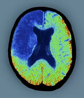
Background imageAbnormal Collection: Cerebral stroke, CT scan

Cerebral stroke, CT scan
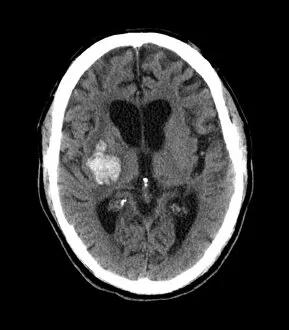
Coloured computed tomography (CT) scan in axial section of the brain of a 65 year old patient showing death (infarction) of cerebral tissue (at upper left) due to a stroke (cerebrovascular accident)

Background imageAbnormal Collection: Pelvis with a leaning posture, X-ray

Pelvis with a leaning posture, X-ray
Coloured X-ray (front view) of the pelvis in a patient with severe knee pain whose walk was affected on one side (at left) and whose posture was lateralised into a sideways leaning gait

Background imageAbnormal Collection: Growth disorder of thigh bone, X-ray

Growth disorder of thigh bone, X-ray
X-ray (front view) of the pelvis in an 8 year old patient showing ossification nuclei in the head of the thigh bone (femur, at left) due to the bone growth disorder chondrodysplasia punctata

Background imageAbnormal Collection: Collapse (atelectasis) of lung, X-ray

Collapse (atelectasis) of lung, X-ray
Coloured X-ray (front view) of the chest in a 58 year old patient showing collapse (atelectasis) of part of the upper lobe of a lung (orange, at left)

Background imageAbnormal Collection: Lung abscess, CT scan

Lung abscess, CT scan
Coloured computed tomography (CT) scan in axial view of the lung fields in a 47 year old patient showing the presence of an abscess (black) in each lung

Background imageAbnormal Collection: Sprained ankle, X-ray

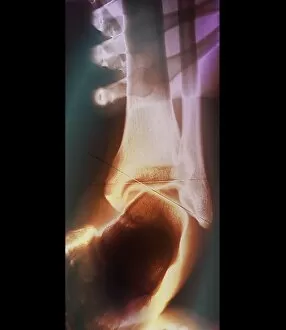
Sprained ankle, X-ray
Coloured X-ray (posterior view) of the sprained ankle in a 25 year old footballer showing the sideways angulation of the foot in relation to the leg due to overstretching the joints ligaments

Background imageAbnormal Collection: Slipped end of thigh bone, X-ray

Slipped end of thigh bone, X-ray
Coloured X-rays of the right hip of a young patient showing the slipped end (epiphysis) of the thigh bone (femur). During childhood the ends of the long bones are busy growing

Background imageAbnormal Collection: Fusion of spinal bones, X-ray

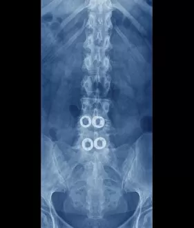
Fusion of spinal bones, X-ray
Coloured X-ray (front view) of the spine in a 45 year old patient with degenerative disc disease showing two implants (prostheses) between vertebral bones L4-L5 and L5-S1

Background imageAbnormal Collection: Arthritic hands

Arthritic hands
MODEL RELEASED. Arthritic hands. Hands of a person that have become deformed due to arthritis. Arthritis is a degenerative condition that affects the joints causing inflammation and pain

Background imageAbnormal Collection: Prostate disorder, X-ray urogram

Prostate disorder, X-ray urogram
Coloured X-ray urogram of the pelvis in a 63 year old male patient showing the bladder (light) with the enlarged prostate gland beneath

Background imageAbnormal Collection: Bone growth disorder of skull, X-ray

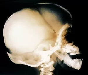
Bone growth disorder of skull, X-ray
Coloured X-ray (side view) of the skull in a 7 year old patient with bone growth abnormalities including wide-open sutures to the fontanelle

Background imageAbnormal Collection: Atherosclerosis in femoral arteries X-ray

Atherosclerosis in femoral arteries X-ray
Coloured X-ray (front view) of the pelvic region in a 72 year old patient showing atherosclerosis (mottled appearance) in the femoral arteries running on the inside of the femur (thigh) bones

Background imageAbnormal Collection: Spine in Morquio syndrome, X-ray

Spine in Morquio syndrome, X-ray
Coloured X-ray (side view) of the spine in a 7 year old patient with Morquio syndrome showing deformed vertebral bones (osteodysplasia). Morquio syndrome is an inherited lysosomal storage disorder

Background imageAbnormal Collection: Dolichocephalic skull deformity, X-ray

Dolichocephalic skull deformity, X-ray
Coloured X-ray (side view) of the elongated skull (dolichocephalic deformity) in an adult patient. This deformed skull can either characterize newborns who are born after breech presentation

Background imageAbnormal Collection: Rheumatoid arthritis of the hip, X-ray

Rheumatoid arthritis of the hip, X-ray
Coloured X-ray (front view) of the pelvis in a 58 year old patient showing rheumatoid arthritis of the hip joint (at right)

Background imageAbnormal Collection: Fibrosis on lower leg, X-ray

Fibrosis on lower leg, X-ray
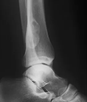
Fibrosis on lower leg. X-ray (side view) of the right ankle of a patient showing fibrosis (scar tissue, upper centre) at the inferior (lower) end of the tibia (shin bone)

Background imageAbnormal Collection: Sickle cell anaemia, artwork

Sickle cell anaemia, artwork
Sickle cell anaemia. Computer artwork of normal red blood cells (erythrocytes, red) and red blood cells affected by sickle cell anaemia (pink)

Background imageAbnormal Collection: Cystic pancreas tumour, CT scan

Cystic pancreas tumour, CT scan
Computed tomography (CT) scan in axial view through the abdomen in a 60 year old patient showing a large adenoma (benign tumour) on the head of the pancreas (grey, upper left)

Background imageAbnormal Collection: Dislocated finger joint, X-ray

Dislocated finger joint, X-ray
Coloured X-ray (two views) of the dislocation (subluxation) of a finger joint (interphalangeal joint) after an injury

Background imageAbnormal Collection: Familial hypercholesterolaemia, diagram

Familial hypercholesterolaemia, diagram
Familial hypercholesterolaemia. Diagram showing the biochemical processes that take place in a case of the genetic disorder familial hypercholesterolaemia

Background imageAbnormal Collection: Subarachnoid haemorrhage, MRI scan

Subarachnoid haemorrhage, MRI scan
Subarachnoid haemorrhage. Magnetic resonance imaging (MRI) scan of an axial section through the brain of a patient with a subarachnoid haematoma (light grey, centre left)

Background imageAbnormal Collection: Berry aneurysm, angiogram

Berry aneurysm, angiogram
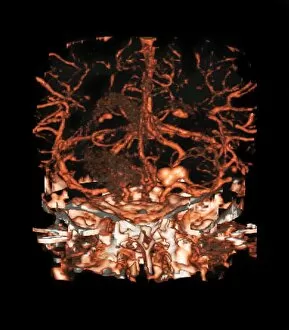
Berry aneurysm. Coloured angiogram of the brain of a patient with an aneurysm (lower centre right) in a cerebral artery. This type of aneurysm is known as a berry, or saccular, aneurysm

Background imageAbnormal Collection: Hip fracture, digital X-ray

Hip fracture, digital X-ray
Hip fracture. Digital frontal X-ray of the pelvis and hips of a patient in their mid eighties. This patient has a fracture of the neck of their left femur (upper leg bone)

Background imageAbnormal Collection: Male Downs syndrome karyotype, artwork

Male Downs syndrome karyotype, artwork
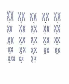
Male Downs syndrome karyotype, artowrk. The complete set of chromosomes of an organism is known as the karyotype. Humans usually have 46 chromosomes in total, arranged into 23 pairs

Background imageAbnormal Collection: Balloon angioplasty, artwork

Balloon angioplasty, artwork
Balloon angioplasty. Artwork showing balloon angioplasty treatment for stenosis (narrowing) of a coronary artery. A balloon catheter (blue)

Background imageAbnormal Collection: Angioplasty, artwork

Angioplasty, artwork
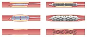
Angioplasty. Artwork showing two treatments for stenosis (narrowing) of arteries. At left is balloon angioplasty, where a balloon catheter is inserted into the affected artery

Background imageAbnormal Collection: Asthma pathology, artwork

Asthma pathology, artwork
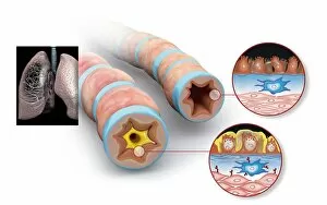
Asthma pathology. Computer artwork showing cross-sections through a normal airway (right) and an asthmatic, constricted airway (left)

Background imageAbnormal Collection: Elbow replacement, artwork

Elbow replacement, artwork
Elbow replacement. Artwork showing a prosthetic elbow joint in place. The elbow joint is formed by the articulation of the humerus (upper arm bone) and the ulna and radius (lower arm bones)

Background imageAbnormal Collection: Vascular disease progression, artwork

Vascular disease progression, artwork
Vascular disease progression. Artwork showing the progression of vascular disease in cross-section (left) and transverse section (right). At top is a healthy artery

Background imageAbnormal Collection: Narrowed blood vessel, artwork

Narrowed blood vessel, artwork
Narrowed blood vessel. Cut-away artwork showing a blood vessel that is partially blocked by atheroma. Atheroma is the deposition of fatty material on the inner walls of a blood vessel

Background imageAbnormal Collection: Amputated fingertip, X-ray

Amputated fingertip, X-ray
Amputated fingertip. Coloured X-ray of the 4th and 5th fingers of a hand where the end of the 5th (little) finger has been amputated (red)

Background imageAbnormal Collection: Gallbladder and biliary tree, 3D MRI

Gallbladder and biliary tree, 3D MRI
Gallbladder and biliary tree. Three-dimensional magnetic resonance image (MRI) showing the gallbladder (round) and biliary tract (branching)

Background imageAbnormal Collection: Ingrowing toenail, artwork

Ingrowing toenail, artwork
Ingrowing toenail. Artwork showing a normal toenail (bottom left) and an ingrowing toenail (top and bottom right). An ingrowing toenail is deformed and grows into the flesh of the toe

Background imageAbnormal Collection: Glaucoma, artwork

Glaucoma, artwork
Glaucoma. Artwork showing glaucoma arising from the drainage system of the eye becoming blocked (bottom right) and a normal eye (top right)

Background imageAbnormal Collection: Emphysema, artwork

Emphysema, artwork
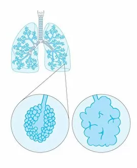
Emphysema. Artwork showing the lungs with a detailed view of a normal alveolus (air sac, left) and an alveolus affected by emphysema (right)

Background imageAbnormal Collection: Endometriosis, artwork

Endometriosis, artwork
Endometriosis. Artwork of a section through the female pelvis, showing the movement of cells from the lining of the uterus to the abdominal cavity in endometriosis

Background imageAbnormal Collection: Kidney stone, artwork

Kidney stone, artwork
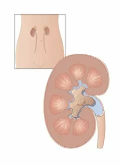
Kidney stone. Artwork of a large kidney stone. Kidney stones are formed when mineral salts, most often involving calcium, accumulate on the inner surfaces of the kidney

Background imageAbnormal Collection: Foot fractures, artwork

Foot fractures, artwork
Foot fractures. Artwork showing a healthy foot (top), a foot with a fractured metatarsal (bottom right) and a foot with a fractured calcaneus (heel) bone (bottom left)

Background imageAbnormal Collection: Applying eye drops, artwork

Applying eye drops, artwork
Applying eye drops. Artwork showing how to apply eye drops to an itchy eye


For sale as Licensed Images

Choose your image, Select your licence and Download the media

"Unveiling the Abnormal: A Glimpse into Extraordinary Phenomena" Gas pillars in the Eagle Nebula: Witness the mesmerizing dance of celestial gas pillars, defying gravity and painting a surreal picture in the depths of space. Apollo 11 astronaut footprint on Moon: Behold an iconic abnormality, a single footprint etched by humanity's giant leap onto another world, forever marking our presence among the stars. Broken wrist bone, X-ray C017 / 7187: Delve into the intricate realm of human anatomy as we explore an abnormality captured within an X-ray image – a fractured wrist bone telling tales of resilience and healing. Solar prominence: Marvel at nature's fiery spectacle as solar prominences erupt from our nearest star, showcasing their abnormal beauty against the backdrop of infinite cosmic expanse. DAF 95 XF wide: Discover automotive innovation with this extraordinary truck model boasting exceptional width – its bold design challenges conventional norms to redefine road transport. HeLa cells, light micrograph C017 / 8299: Peer through a microscope lens to witness HeLa cells – immortalized yet controversial entities that have revolutionized medical research while raising ethical questions about their origin. Image of Jupiter taken with Hubble Telescope: Explore Jupiter's enigmatic atmosphere through an extraordinary image captured by Hubble Telescope; revealing swirling storms and vibrant bands that defy expectations. Knee pain, conceptual artwork: Dive into thought-provoking artistry depicting knee pain – a visual representation inviting contemplation on physical discomforts experienced by many but often hidden beneath societal masks. Joint pain, conceptual artwork: Engage your senses with evocative imagery portraying joint pain; transcending boundaries between art and reality to shed light on invisible struggles faced by individuals worldwide. Total hip replacement, X-ray.