Alga Collection (#18)

"Discovering the Intricate Beauty of Algae: From Ernst Haeckel's Art to 19th-Century Cyanotypes and SEM Images" Immerse yourself in the captivating world of algae

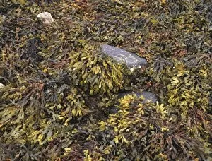
Background imageAlga Collection: Bladder wrack seaweed

Bladder wrack seaweed (Fucus vesiculosus) thallus in a rocky shore. The name of the seaweed derives from the paired air bladders visible on either side of the midrib of the fronds

Background imageAlga Collection: Blue-green alga, Spirulina platensis

Blue-green alga, Spirulina platensis
False colour transmission electron micrograph of the blue-green algae, Spirulina platensis. Here, a single cell of this species is seen. Blue-green algae (cyanobacteria) are primitive algae

Background imageAlga Collection: Periphyton algae (Periphyton sp. )

Periphyton algae (Periphyton sp. )
Periphyton algae (Periphyton sp.) floating on the water surface. Periphyton is a complex matrix of algae, cyanobacteria (blue-green algae), heterotrophic microbes

Background imageAlga Collection: Seaweed

Seaweed

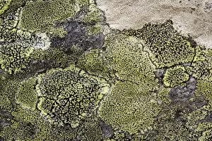
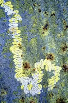
Background imageAlga Collection: Map lichen (Rhizocarpon geographicum)

Map lichen (Rhizocarpon geographicum) on a rock. Lichens are symbiotic organisms formed of a fungus and an alga that mutually benefit one another. Photographed in Scotland

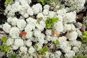
Background imageAlga Collection: Lichen (Cladonia stellaris)

Lichen (Cladonia stellaris)
Cladonia stellaris lichen (white) and lingonberry (Vaccinium vitis-idaea, green and orange). Lichens are symbiotic organisms formed of a fungus and an alga that mutually benefit one another

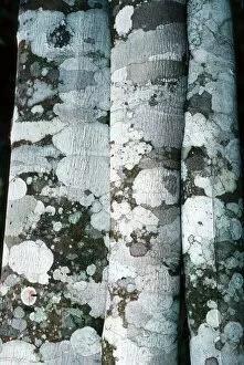
Background imageAlga Collection: Lichen on cinnamon trees

Lichen on cinnamon trees
Lichen on cinnamon tree trunks (Cinnamomum verum). Lichens are formed from the symbiotic association between a fungus and an alga. The alga is contained within the hyphae (filaments) of the fungus

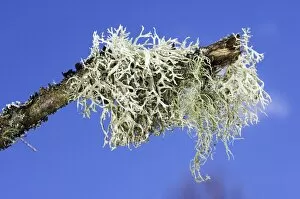
Background imageAlga Collection: Lichen (Hypogymnia physodes)

Lichen (Hypogymnia physodes) on a twig. Lichens are formed from the a symbiotic association between a fungus and an alga. The alga is contained within the hyphae (filaments) of the fungus

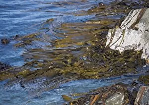
Background imageAlga Collection: Kelp seaweed

Kelp seaweed (Laminaria digitata) floating at the waters surface. This seaweed is also known as devils apron and oarweed. It is used in the production of iodine, soda and fertilisers

Background imageAlga Collection: Seaweed covered beach

Seaweed covered beach. These are mostly kelps, tangles and oar weeds. There is a covering of several feet after the seaweed was washed on to the beach after a storm

Background imageAlga Collection: Furbelows seaweed, Saccorhiza polyschides

Furbelows seaweed, Saccorhiza polyschides
Furbelows seaweed holdfast (Saccorhiza polyschides). This part of the plant anchors to the rock. Furbelows is the largest European seaweed, it can grow up to 4.5 metres in length

Background imageAlga Collection: Bladder wrack (Fucus vesiculosus)

Bladder wrack (Fucus vesiculosus) seaweed in a rock pool. As a good source of iodine this seaweed was used in the nineteenth century to treat goitre

Background imageAlga Collection: Knotted wrack (Ascophyllum nodosum)

Knotted wrack (Ascophyllum nodosum) seaweed in a rock pool. This seaweed is found in many coastal habitats of the northern Atlantic Ocean

Background imageAlga Collection: Bull kelp bed

Bull kelp bed (Durvillea antarctica) near Kaikoura, South Island, New Zealand

Background imageAlga Collection: Common stonewort (Chara vulgaris)

Common stonewort (Chara vulgaris). Photographed in Greece, in April

Background imageAlga Collection: Marine diatom alga, SEM

Marine diatom alga, SEM
Diatom alga. Coloured scanning electron micrograph (SEM) of part of a marine diatom alga. Diatoms form a large part of the marine phytoplankton

Background imageAlga Collection: Chlamydomonas alga

Chlamydomonas alga
Coloured Transmission Electron Micrograph of a section through the unicellular green algae, Chlamydomonas asymmetrica. The micrograph shows the arrangement of chloroplasts (green)

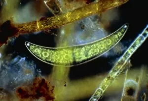
Background imageAlga Collection: Closterium, a desmid alga

Closterium, a desmid alga

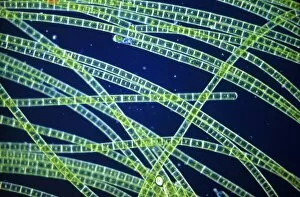
Background imageAlga Collection: Green filamentous alga, Zygnema

Green filamentous alga, Zygnema
Light micrograph of the common green filamentous alga Zygnema. Magnification: x28 at 35 mm size

Background imageAlga Collection: Algae

Algae. Partly eaten algae on a rock. The tracks of a molluscs radula (rasping tongue) can be seen

Background imageAlga Collection: Freshwater alga, SEM

Freshwater alga, SEM
Freshwater alga. Coloured scanning electron micrograph (SEM) of a single-celled freshwater alga

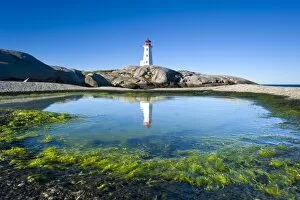
Background imageAlga Collection: Peggys Cove, Canada

Peggys Cove, Canada
Peggys cove. Lighthouse, tide pool and marine algae at Peggys point in Peggys cove, Nova Scotia, Canada

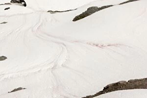
Background imageAlga Collection: Red snow, / Watermelon snow, caused by unicellular algae (Chlamydomonas nivalis)

Red snow, / Watermelon snow, caused by unicellular algae (Chlamydomonas nivalis), on Mount Rainier, Cascade Mountains
ROG-12941 Red snow / Watermelon snow, caused by unicellular algae on Mount Rainier, Cascade Mountains, Washington Chlamydomonas nivalis Bob Gibbons Please note that prints are for personal display


For sale as Licensed Images

Choose your image, Select your licence and Download the media

"Discovering the Intricate Beauty of Algae: From Ernst Haeckel's Art to 19th-Century Cyanotypes and SEM Images" Immerse yourself in the captivating world of algae, as we delve into its mesmerizing artistry and scientific wonders. Ernst Haeckel, a renowned biologist and artist, showcases the delicate intricacy of diatom algae through his stunning illustrations. These masterpieces reveal the symmetrical beauty hidden within these microscopic organisms. Step back in time with pressed seaweed specimens C016 / 6127 from centuries ago. Preserved meticulously, they offer a glimpse into the diverse forms and colors that algae can take on. The cyanotype technique used during the 19th century captures their essence in ethereal blue hues, adding an enchanting touch to their already fascinating nature. Calcareous phytoplankton observed under scanning electron microscopy (SEM) unravels another layer of algal marvels. Discosphaera tubifera, a coccolithophore species adorned with intricate calcium carbonate plates, showcases nature's architectural genius at work. Dictyota dichotoma emerges as a striking example of brown algae thriving along coastlines worldwide. Its branching structure creates an artistic spectacle underwater while providing essential habitats for marine life. Diatoms continue to amaze us with their geometric patterns when viewed under SEM imaging techniques. Their microscopic beauty is unveiled through meticulous detailing that reveals their remarkable diversity and ecological significance. Venturing further into aquatic realms, Fucus bulbosus or kelp stands tall as one of nature's giants beneath ocean waves. This majestic alga plays a crucial role in coastal ecosystems by offering shelter to numerous marine organisms. Pond life surprises us with its vibrant assortment of microorganisms where diatoms once again steal the show under SEM imaging techniques. Their intricate designs resemble miniature works of art floating amidst water bodies' tranquility.