Anatomical Collection (page 33)

"Unveiling the Anatomical Wonders: Exploring the Intricacies of Life" Step into a world where science meets art, as we delve into the fascinating realm of anatomy

Background imageAnatomical Collection: Wood engraving

Wood engraving
HUMAN HEART. Wood engraving

Background imageAnatomical Collection: Aer-vessels (i.e. vascular bundles) unrooved in a vine leaf. Line engraving from The Anatomy of

Aer-vessels (i.e. vascular bundles) unrooved in a vine leaf. Line engraving from The Anatomy of Plants
GREW: PLANT ANATOMY, 1682. Aer-vessels (i.e. vascular bundles) unrooved in a vine leaf. Line engraving from The Anatomy of Plants... (1682), by Nehemiah Grew (1641-1721)

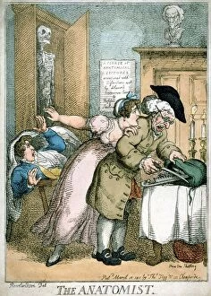
Background imageAnatomical Collection: The Anatomist cartoon by Rowlandson published 1811, showing terror of patient

The Anatomist cartoon by Rowlandson published 1811, showing terror of patient and wife as surgeon opens bag of instruments

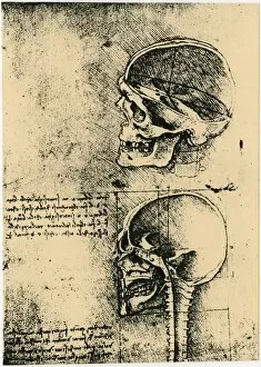
Background imageAnatomical Collection: Leonardo da Vinci (1452-1519) Italian painter, sculptor, engineer, architect. Anatomical

Leonardo da Vinci (1452-1519) Italian painter, sculptor, engineer, architect. Anatomical drawings of a skull in profile with cranium removed

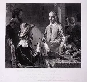
Background imageAnatomical Collection: Harvey demonstrating circulation of the blood to Charles I. William Harvey (1578-1657)

Harvey demonstrating circulation of the blood to Charles I. William Harvey (1578-1657), English physician, published his famous De motu cordis

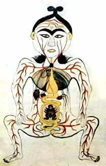
Background imageAnatomical Collection: 17th manuscript (Persian) showing anatomical illustration

17th manuscript (Persian) showing anatomical illustration

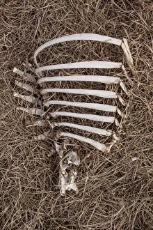
Background imageAnatomical Collection: Red Deer rib cage rotting on Scottish hill side

Red Deer rib cage rotting on Scottish hill side

Background imageAnatomical Collection: Red Deer sculls and bones dumped on Scottish hill side

Red Deer sculls and bones dumped on Scottish hill side

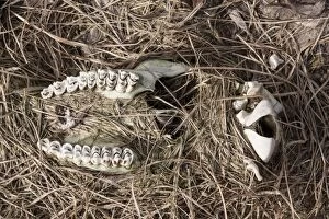
Background imageAnatomical Collection: Red Deer scull showing upper jaw and teeth

Red Deer scull showing upper jaw and teeth

Background imageAnatomical Collection: Model of Spinner Shark (Carcharhinus brevipinna), side view

Model of Spinner Shark (Carcharhinus brevipinna), side view

Background imageAnatomical Collection: Illustration of man with a large goiter on his neck. Lithograph, 1822

Illustration of man with a large goiter on his neck. Lithograph, 1822
MEDICINE: GOITER, 1822. Illustration of man with a large goiter on his neck. Lithograph, 1822

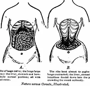
Background imageAnatomical Collection: Two pictures of a womans torso, one showing a woman without a corset

Two pictures of a womans torso, one showing a woman without a corset, the other showing the effects on the internal
NATURE VERSUS CORSETS, 1903. Two pictures of a womans torso, one showing a woman without a corset, the other showing the effects on the internal organs and bones while wearing a corset

Background imageAnatomical Collection: Lepus Cuniculus, Rabbit

Lepus Cuniculus, Rabbit
Plate XVIII of a dissection of a Rabbit from Anatomy of Vertebrates by Daniel McAlpine, published in 1881

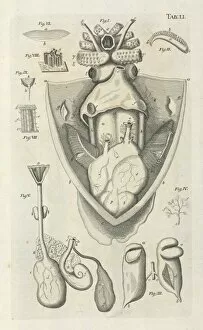
Background imageAnatomical Collection: Cuttlefish dissection drawing

Cuttlefish dissection drawing
Table LI, taken from Bibel der Natur byJan Swammerdamm

Background imageAnatomical Collection: On the dredge

On the dredge
Common dredge as descibed by Professor Edward Forbes. Anatomical Manipulation, Tulk and Henfrey 1843

Background imageAnatomical Collection: Anatomical study of a ray

Anatomical study of a ray
Page 451 by Ulisse Aldrovandi from his De Piscubis et de Cetis, 1638

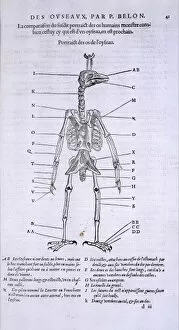
Background imageAnatomical Collection: Skeleton of a bird

Skeleton of a bird
Page 41 from Pierre Belons Histoire de la Nature (1555). Compare with skeleton of a man on page 40, (picture ref. 6886)

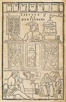
Background imageAnatomical Collection: KETHAM, Johannes de (second half 15th century-begin

KETHAM, Johannes de (second half 15th century-begin
" KETHAM, Johannes de (second half 15th century-begin 16th). Human health compendium" (Fasciculus medicinae, 1491). First medicine book including anatomical illustrations

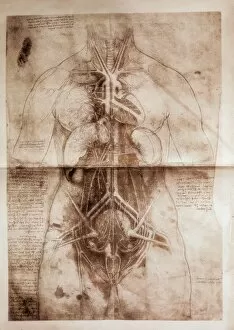
Background imageAnatomical Collection: Anatomical study

Anatomical study
A study of female anatomy by Leonardo Da Vinci (1452-1519)

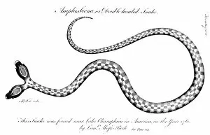
Background imageAnatomical Collection: DOUBLE-HEADED SNAKE, 1764. Snake found near Lake Champlain, 1764, by Lieutenant Moses Park

DOUBLE-HEADED SNAKE, 1764. Snake found near Lake Champlain, 1764, by Lieutenant Moses Park, who drew it. Line engraving from Edward Bancrofts An Essay on the Natural History of Guiana, London, 1769

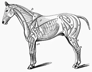
Background imageAnatomical Collection: HORSE: MUSCLE STRUCTURE. Line engraving, 19th century

HORSE: MUSCLE STRUCTURE. Line engraving, 19th century

Background imageAnatomical Collection: Petticoat Lane market doctor

Petticoat Lane market doctor
At the Petticoat Lane market, London, a doctor, dressed as if he was a Harley Street professional, offers advice to passers by with the aid of an anatomical model of a stomach interior. Date: 1945

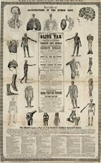
Background imageAnatomical Collection: Anatomical, illustrations of the human body

Anatomical, illustrations of the human body. Date 1857 Southern District of N.Y

Background imageAnatomical Collection: Anatomy School, Oxford

Anatomy School, Oxford
The Anatomy School, Oxford Date: 1821

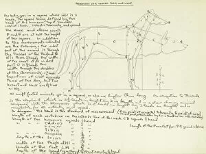
Background imageAnatomical Collection: Proportions of a typical dog and wolf

Proportions of a typical dog and wolf
Detailed anatomical drawing by Raymond Sheppard showing the proportions of a typical dog and wolf, accompanied by copious notes demonstrating Sheppards precise technical interest

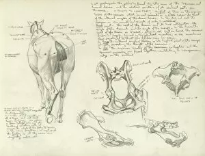
Background imageAnatomical Collection: Studies of a horse

Studies of a horse
Studies of the pelvis of a horse, including a rear view of a horse and annotated studies of individual bones

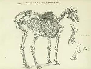
Background imageAnatomical Collection: Skeleton of a horse

Skeleton of a horse drawn at the Natural History Museum, London

Background imageAnatomical Collection: Diagrams of human shoulder and ankle joints

Diagrams of human shoulder and ankle joints
Diagrams of the human shoulder and ankle joints

Background imageAnatomical Collection: Diagram showing the circulation of the blood

Diagram showing the circulation of the blood
A diagram showing the circulation of the blood, with veins, arteries and capillaries

Background imageAnatomical Collection: Gaspar Aselli

Gaspar Aselli
GASPAR ASELLI Italian surgeon from Cremona, practised in Milano, professor at Pavia : made important anatomical discoveries

Background imageAnatomical Collection: First Dissection Pic

First Dissection Pic
This is said to be the earliest depiction of an anatomical dissection

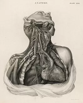
Background imageAnatomical Collection: Dissection of upper torso, showing the heart

Dissection of upper torso, showing the heart
A dissection of the upper torso of a human body, showing the heart, lungs, veins and arteries

Background imageAnatomical Collection: Anatomical Figure 1493

Anatomical Figure 1493
Anatomical figure

Background imageAnatomical Collection: Arteries of the human body

Arteries of the human body
A diagram showing the arteries of the human body


For sale as Licensed Images

Choose your image, Select your licence and Download the media

"Unveiling the Anatomical Wonders: Exploring the Intricacies of Life" Step into a world where science meets art, as we delve into the fascinating realm of anatomy. From Leonardo da Vinci's captivating depiction of a bear's head to his meticulous study of a greyhound's skeleton, these timeless masterpieces offer us glimpses into the intricate workings of living beings. As we journey further, we encounter detailed diagrams showcasing the complexity and beauty of our own bodies. The lungs and bronchial tubes reveal their delicate network, ensuring every breath sustains us. The human brain and spinal column stand as guardians of our thoughts and movements, orchestrating an orchestra within. Our exploration takes us deeper still, unveiling the heart's secrets through cross-sections that expose its innermost chambers. French wood engravings from the late 19th century transport us back in time to witness this vital organ in all its glory. But it is not just our internal structures that captivate; our limbs too hold stories waiting to be told. Diagrams meticulously illustrate each bone in hand and arm or right leg and hip – reminding us that beneath our skin lies an intricate framework supporting every step we take. And finally, a modern twist awaits with an MRI-style X-ray revealing a leg adorned with a stiletto shoe – merging technology with artistry while peering beyond appearances. From ancient manuscripts like "De humani corporis fabrica" to contemporary medical imaging techniques, these anatomical wonders remind us that beneath outward appearances lie marvels waiting to be discovered. They invite us to appreciate both the fragility and resilience encapsulated within each living being – truly celebrating life itself through understanding its intricacies.