
Asexual Collection
"Asexual: Unveiling the Intricate World of Reproduction Through Microscopy" Delving into the microscopic realm


For sale as Licensed Images
Choose your image, Select your licence and Download the media
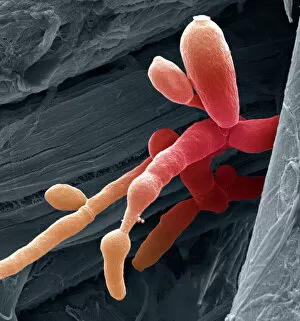
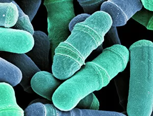
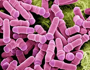
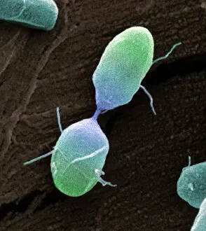
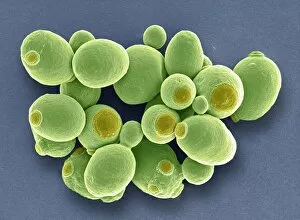
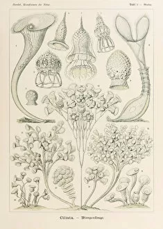
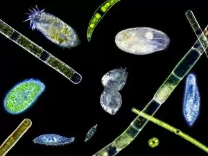
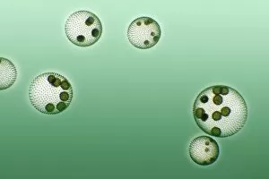
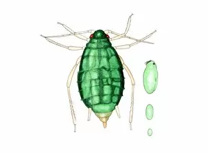
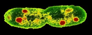
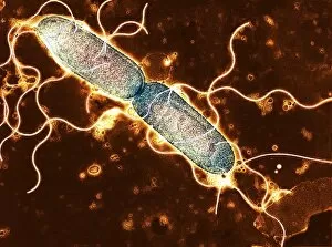
"Asexual: Unveiling the Intricate World of Reproduction Through Microscopy" Delving into the microscopic realm, we encounter a fascinating array of organisms engaging in asexual reproduction. Candida fungus, captured under the scanning electron microscope (SEM), reveals its intricate structure as it proliferates and thrives. Observing dividing yeast cells through SEM provides us with a glimpse into their remarkable ability to reproduce without the need for mating. The process unfolds before our eyes, showcasing nature's ingenuity in perpetuating life. Venturing further, we explore intestinal protozoan parasites using transmission electron microscopy (TEM). These tiny creatures navigate their complex life cycles through asexual means, adapting and surviving within their hosts. The Salmonella bacterium divides under SEM, highlighting its resilience and adaptability. This splitting process showcases how these microorganisms multiply rapidly, posing challenges to human health. Examining bread mold through SEM unravels its intricate network of filaments that aid in spore production. Asexually reproducing by releasing countless spores into the environment ensures this organism's survival and dispersal. Yeast cells come alive under SEM as they undergo division - an essential mechanism for propagation without sexual reproduction. Witnessing this phenomenon reminds us of nature's diverse strategies for continuation. Plate 3 from Ernst Haeckel's "Kunstformen der Natur" introduces Stentor Ciliata – captivating single-celled organisms that engage in various modes of reproduction including asexual methods. Haeckel's illustrations bring forth both scientific accuracy and artistic beauty. Amidst these microscopic wonders lies another form of artistry; Javanese Dancers grace our visual senses with elegant movements symbolizing cultural expressions while reminding us that even humans possess unique ways to propagate life beyond sexuality. Black vine weevil presents itself magnificently under SEM revealing its exoskeleton intricacies while silently multiplying via parthenogenesis – yet another example reproduction in the animal kingdom.